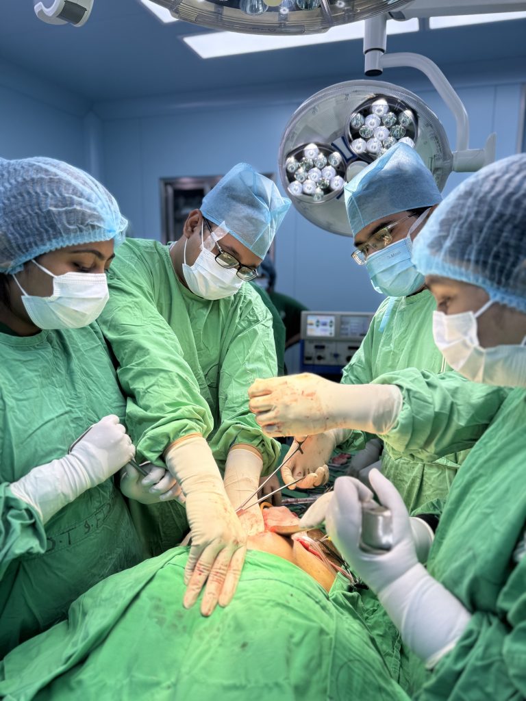
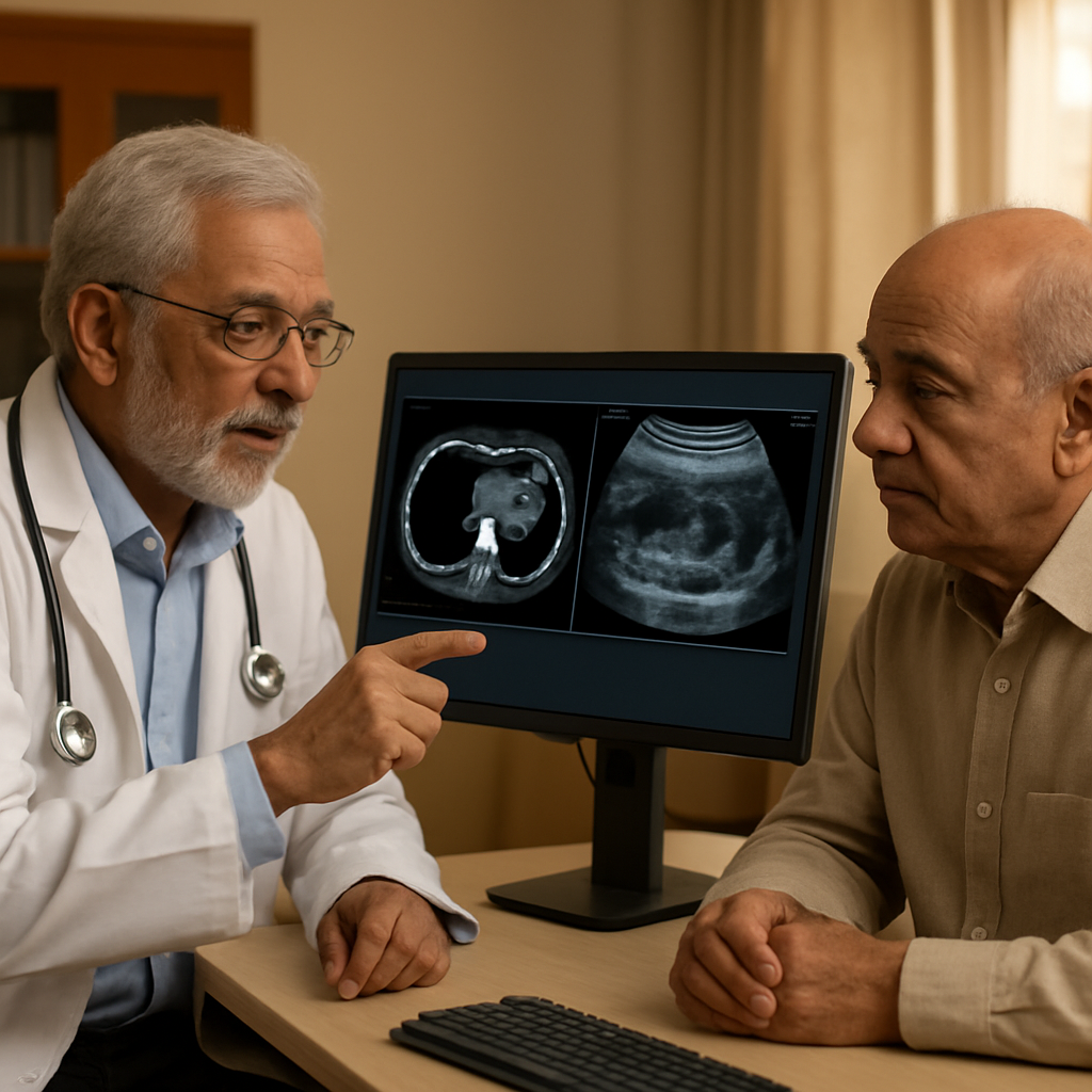
In Dhaka's busy hospitals, a patient's quiet symptoms become a turning point in a pancreatic cancer journey. This post traces the path from early signs and blood tests, including the CA 19-9 test, to imaging, diagnostic tests, and the surgical decision, illustrating how pancreatic cancer early detection can influence outcomes. You'll learn how risk factors, a multidisciplinary care approach, and practical steps—navigating resources, seeking second opinions, and planning recovery—shape what comes next.
Frequently Asked Questions
Key point: In real-world practice, pancreatic cancer early detection remains the exception rather than the rule. Most patients are diagnosed after symptoms appear or when the disease has already progressed, which limits curative options. In Dhaka, access to advanced screening varies by hospital and neighborhood, but awareness of subtle signs can still tilt the odds in a patient’s favor. Persistent upper abdominal discomfort, unplanned weight loss, dark urine or pale stools, and new-onset jaundice are red flags that deserve prompt evaluation, not quiet dismissal. Early discussion with a trusted clinician can shorten the path to imaging and specialist input.
Imaging and tests in practice
Imaging strategy matters: Imaging and tests are not equally useful at every step; the choice depends on risk, symptoms, and local availability. In Dhaka’s clinics, ultrasound is typically the first screen because it’s quick and accessible, but it can miss small tumors or those in certain parts of the pancreas. If suspicion remains, contrast-enhanced CT is usually the next anchor test for characterization and staging. MRI offers greater soft-tissue detail when needed, and endoscopic ultrasound (EUS) provides the highest sensitivity for tiny lesions, though it requires referral to a specialized center. The CA 19-9 blood marker helps with monitoring known cancer or tracking response, but it is not a reliable screening tool and can be elevated for benign conditions.
Concrete example: A 58-year-old patient in a Dhaka private hospital presents with weight loss and mild jaundice. An ultrasound flags a suspicious pancreatic lesion; a contrast CT confirms a mass, and the patient is fast-tracked to a multidisciplinary tumor board. Because the team moves quickly, she starts surgical planning before the disease advances, illustrating how timely imaging and coordinated care changes outcomes in practice.
Important caveat: A negative CA 19-9 does not rule out cancer, and elevated CA 19-9 can occur with non-cancer conditions. Do not rely on a single test for diagnosis. Use CA 19-9 as a supplementary piece of the puzzle when there is already clinical suspicion or a confirmed diagnosis, and always interpret results in the context of imaging and symptoms.
Next steps for readers in Dhaka: If risk factors exist, request a formal assessment with a gastroenterologist and an oncologist. Start with your primary care physician to map a plan, clarify imaging options (ultrasound, CT, MRI, EUS), discuss costs and access, and arrange a second opinion at a center with a dedicated pancreatic team. Keep a simple symptom log and bring it to every visit.
Related Articles

How to Prepare for Upper GI Surgery: A Step-by-Step Guide for Patients at Popular Medical College Hospital
How to Prepare for Upper GI Surgery: A Step-by-Step Guide for Patients at Popular Medical College Hospital Preparation for upper […]
Read More
Surgical Baseline Assessment at Entry to Training
Surgical Baseline Assessment at Entry to HPB Training in Dhaka Most trainees assume the entry test exists to reject them, […]
Read More
Hepatobiliary Surgery for Liver, Bile Duct and Pancreas Disease
Hepatobiliary Surgery for Liver, Bile Duct and Pancreas Disease Hepatobiliary surgery treats the liver, the bile ducts and the pancreas […]
Read More