
Minimally Invasive Liver Surgery | Dr. Arefin

Minimally Invasive Liver Surgery | Dr. Arefin

Minimally Invasive Liver Surgery for Complex Cases in Dhaka

Keyhole surgery changes the wound, not the liver. The incisions are smaller, the operation inside is the same operation, and a laparoscopic resection that has to be hurried is worse than an open one done calmly.

When a Complex Liver Case Can Still Be Done by Keyhole

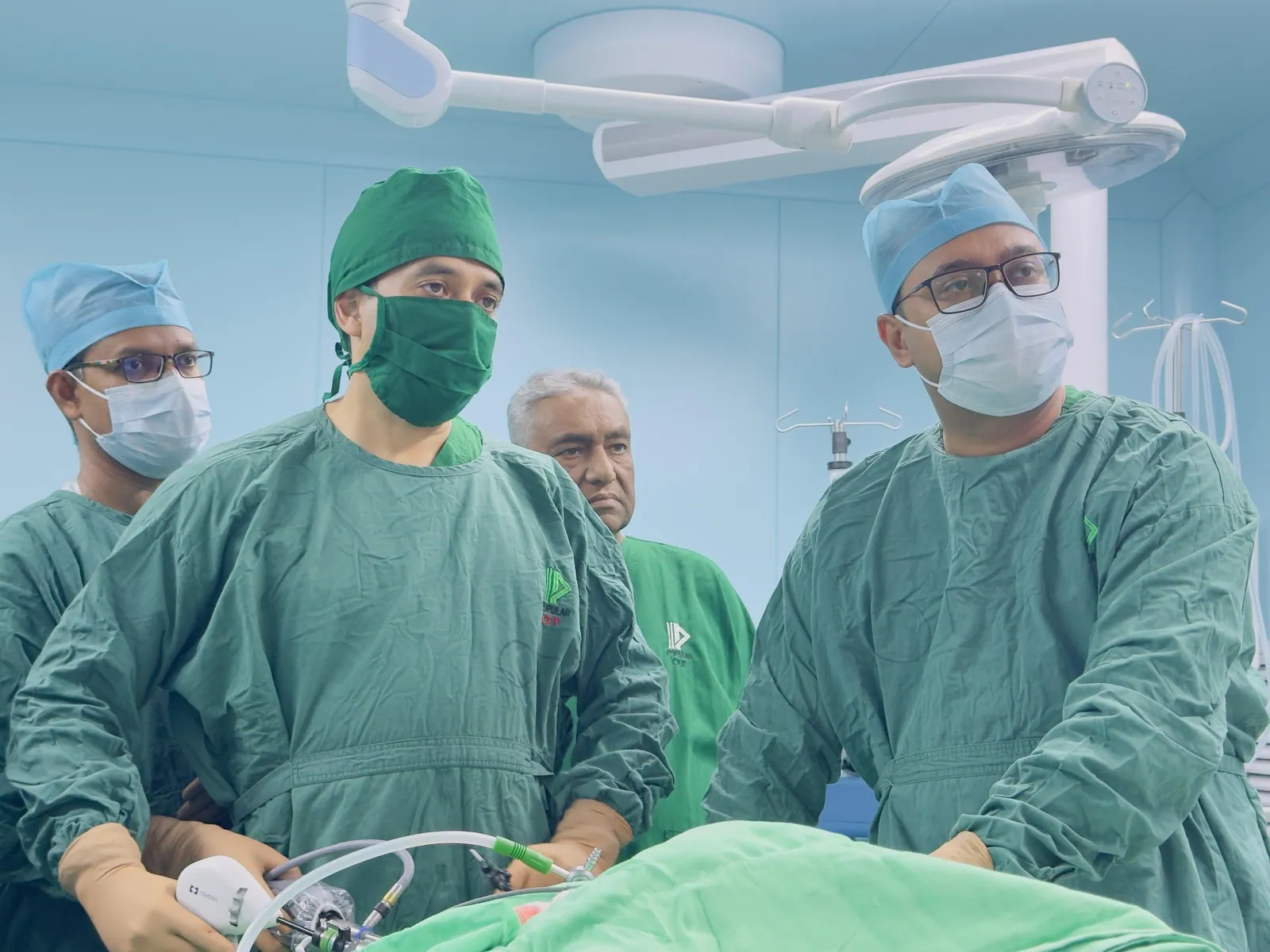
Laparoscopy has moved a long way into liver surgery without replacing open surgery, and whether a case suits it is decided by the scans, not by preference. Lesions in the left lateral segments, peripheral lesions in the anterior right segments, wedge resections, left lateral sectionectomy, deroofing of a symptomatic cyst and staging before a bigger operation suit minimally invasive liver surgery most comfortably. Selected right-sided and major resections are done this way in experienced hands.

Three conditions have to hold first. The oncological result must be unchanged, meaning the same clearance and margin an open operation would give. Bleeding must be controllable through the ports, with inflow control prepared before the parenchyma is divided. And conversion to open surgery must be available at any moment without it feeling like a crisis, which means the patient is consented for both and the theatre is set up for both. Where any of those is in doubt, the correct operation is open. A patient with an intact liver and a longer scar is in a far better position than one with a short scar and an avoidable complication.

Key Takeaways on Minimally Invasive Liver Surgery:

  • Keyhole access improves the recovery from the wound. It does not change the liver’s tolerance of resection.
  • Peripheral and left-sided lesions suit laparoscopy best; posterosuperior segments, caval and hilar proximity and dense adhesions push towards open surgery.
  • The same volumetric and liver function assessment applies whichever approach is used.
  • Conversion to open surgery is a planned option, decided on judgement, and is not a failure of the operation.
  • Major bile duct reconstruction is usually done open, even in units that do most gallbladder and liver work laparoscopically.

Tumour Position, Size and Vessels That Change the Plan

Position matters more than size. A lesion in segments 7 or 8, high and behind, is hard to control through ports, and reaching it can require mobilising the liver so completely that the advantage disappears. Proximity to the inferior vena cava or the hepatic vein confluence is the other common reason to open, because the manoeuvre that saves a patient from sudden venous bleeding needs a hand in the abdomen. Tumours reaching the hilum need reconstruction, and reconstruction is open work.

The abdomen’s history counts too. Previous upper abdominal surgery or repeated biliary infection leaves adhesions that turn a planned keyhole start into a slow dissection. Cirrhosis with portal hypertension brings a fragile liver, collateral veins in the abdominal wall and poor clotting, and while laparoscopy has real advantages there, it has less margin for error. A very large tumour sometimes needs an incision for extraction anyway, which is better said in the consultation than afterwards.

Families in Bangladesh usually come with the phrase লিভারের অপারেশন and one question underneath it: will the abdomen be opened. The plan is written from the scans, the approach is stated in the consent, and the surgeon reserves the right to change it if the anatomy says so. A complex hepatic resection can be attempted through ports in the right case and abandoned in the wrong one, on the same principle.

A Conversion That Was the Right Call

A man in his forties was listed for a laparoscopic resection of a peripheral right lobe lesion.

He had had two previous operations and the adhesions were worse than the scans suggested. Two hours in, the lesion turned out to sit closer to a major hepatic vein branch than the imaging had implied, so a right subcostal incision was made. The resection was completed with the vein controlled properly, blood loss stayed modest, and he went home on the sixth day rather than the fourth. He asked whether the first two hours had been wasted. They had not: they established that the operation needed a different approach. Shared with permission, identifying details removed.

A South Asian surgeon in a consultation room in Dhanmondi reviewing a triple-phase CT scan for minimally invasive liver surge
Preoperative planning involves a detailed review of CT and MRI scans to determine if a case is suitable for a minimally invasive approach.

Preoperative Liver Function and Volumetric Assessment

The approach does not soften the requirements that come before it. A triple-phase CT or contrast MRI maps the lesion against the portal and hepatic veins, and where a major resection is planned the remnant volume is calculated rather than estimated. Function is assessed separately, because a remnant that looks adequate on the scan can still belong to a fibrotic liver. Bilirubin, albumin, prothrombin time, platelet count, viral serology and nutrition all count, alongside Child-Turcotte-Pugh and the albumin-bilirubin score.

Two points plainly. None of these numbers changes because the operation is done through ports: a liver that cannot tolerate the loss of a lobe will not tolerate it any better through five small incisions. And if the remnant is too small, the answer is portal vein embolisation, a staged operation or a non-surgical treatment, not a smaller wound. That assessment is set out on the complex hepatic resection page and is the same for both approaches. Liver-disease guidance from EASL follows the same reasoning.

Theatre Setup That Makes Keyhole Liver Work Safe

Laparoscopic liver surgery is a joint operation between surgeon and anaesthetist, and most of what keeps it safe is decided before the first port goes in. Central venous pressure is kept low so the hepatic veins are not distended during transection, balanced against the risk of air being drawn into an open vein. Insufflation pressure is set high enough to hold the working space and tamponade small venous bleeding, low enough not to compromise the circulation.

Cardiac and respiratory fitness carry slightly more weight here, because a long pneumoperitoneum raises abdominal pressure and alters how the heart fills, and in patients with poor cardiac reserve that occasionally decides the approach on its own.

The instruments matter as much as the pressures. Laparoscopic ultrasound confirms the lesion and its relation to the vessels, because the surgeon cannot palpate. Energy devices, a vessel sealer and vascular staplers are prepared before transection. A sling for the hepatic pedicle, so inflow can be clamped in seconds, is placed at the outset rather than fetched in an emergency, and an open instrument tray stays ready in theatre. For laparoscopic liver surgery Dhaka patients, the question to ask a unit is not whether it can do keyhole liver work, but whether it is equipped and staffed for the moment a case turns. Society material from SAGES sets out the same standards.

Inside a Laparoscopic Hepatic Resection, Step by Step

Access and ports. The abdomen is entered carefully, more carefully still after previous surgery, and four or five ports are placed to give the camera and both hands a working angle. Old adhesions are divided first.

Assessment. The liver and the whole abdomen are checked for disease the scans did not show. Ultrasound confirms the lesion, looks for others and marks the line of transection.

Mobilisation and control. The liver is freed as far as the resection requires and the pedicle sling is positioned. For an anatomical resection the inflow is identified and divided, and the demarcation on the surface confirms the plan.

Transection. The parenchyma is divided along the marked line with central venous pressure kept low. Vessels and ducts are sealed, clipped or stapled as they are met, and larger hepatic vein branches are taken with a stapler. This is the slow part and it is not hurried.

Retrieval and check. The specimen goes into a retrieval bag and out through an enlarged port site or a small extraction incision, with the margin examined immediately. The cut surface is inspected under low insufflation pressure for bleeding and bile, because a bile leak found now is a dressing change and one found on the fourth day is a readmission. A drain is placed selectively, most often after resections close to the bile ducts.

What Is Done Laparoscopically in Biliary Work Too

Most of the biliary workload is laparoscopic and has been for years. Routine gallbladder removal is keyhole work, and so is the difficult gallbladder: thick-walled, contracted and stuck to the duodenum and colon, where the safe move is often a subtotal cholecystectomy leaving a small cuff rather than a heroic dissection at the neck. Acute cholecystitis is operated laparoscopically too, and timing matters more than access, because an inflamed gallbladder handled early is easier than one handled when the tissues are at their worst.

Bile duct stones are cleared by endoscopy in most patients, with the gallbladder removed afterwards. Where endoscopy cannot reach them, laparoscopic bile duct exploration is possible in selected cases with a choledochoscope.

What is not usually keyhole work is reconstruction. A bile duct injury needing a hepaticojejunostomy, a benign stricture, a choledochal cyst excision, a hilar tumour needing duct resection: these are open operations in most hands, because the anastomosis has to be accurate, unhurried and made on healthy duct, and that decides whether the repair holds for decades. Units offering keyhole liver surgery Bangladesh patients ask for should still be judged on their open reconstructive work, because that is the harder skill.

Less Pain, Shorter Stay: Realistic Recovery Expectations

The benefits are real and specific. Wound pain is less, so breathing is easier and mobilising on the first day is realistic. Bowel function returns sooner and hospital stay is typically shorter by a day or two. Wound infection is less frequent and incisional hernia later is less common, a benefit that only becomes visible a year or two afterwards.

A plain picture of the days afterwards: the night of surgery is spent in a monitored bed with observations, blood tests and pain relief through a drip. Day one brings sitting out, fluids and a walk to the door. Days two and three bring solid food, removal of the catheter and liver function trending back towards normal. Discharge is usually between day three and day five for a minor or peripheral resection, later for a major one or where a drain is still draining. Shoulder tip pain from the gas is common for two or three days and is not a complication.

What does not change deserves equal clarity. The risk of post-hepatectomy liver failure is set by the remnant and its function, not by the size of the incisions. The biology of the cancer is unchanged, so margin, node status and response to chemotherapy decide the long-term outcome, and surveillance runs on the same schedule. Bile leak, collection, bleeding and chest infection all still happen. A patient who goes home on day three has had a better recovery, not a different disease.

Peer-Reviewed Liver Resection Risk Research

The risk assessment behind these operations is grounded in outcome data, including work Dr. Arefin has co-authored. A study of liver resection patients published in Cureus, reported here in the authors’ own figures, found complications in 42.9% of the 30 patients analysed from a cohort of 70. Post-hepatectomy liver failure occurred in 0.0% of patients with ALBI grade 1, 38.5% with ALBI grade 2 and 100% with ALBI grade 3. The preoperative albumin-bilirubin score predicted complications with an area under the curve of 0.659, and at a cutoff of -2.67 gave a sensitivity of 70.0% and a specificity of 62.5%. The full paper is the Cureus research on the preoperative ALBI score and complications after liver resection.

Two honest qualifications. An area under the curve of 0.659 is weak to moderate discrimination, so the score sorts groups better than it predicts individuals and is used as an adjunct to volumetry, Child-Turcotte-Pugh, imaging and clinical assessment rather than as a decision on its own. Dr. Arefin is a co-author of this paper, not its first author.

A third point matters specifically here. That study is about liver function and complications. It says nothing about laparoscopic versus open access and must not be read as evidence that keyhole surgery is safer. The case for the approach rests on wound-related recovery, and the case for operating at all rests on the liver.

“Laparoscopy is an operation performed in the abdomen or pelvis using small incisions with the aid of a camera.”

Source: Laparoscopy, Wikipedia

Getting Your Scans Reviewed Before Surgery

Bring the images, not only the reports. A CT or MRI on a disc or a shared link allows the lesion to be placed against the vessels, which is what decides whether ports are reasonable. Bring previous operation notes, because an old open cholecystectomy changes the expected adhesions, along with any biopsy result, a record of chemotherapy, viral serology with current treatment, and recent bilirubin, albumin, INR, platelet count and creatinine. Mention any cardiac or respiratory history early, because a long pneumoperitoneum is not neutral for the heart. The most useful extra is a list of what you have already been told.

Research-based practice: what Dr. Arefin has published

Dr. Murshidul Arefin is a research-active hepatobiliary surgeon, not only an operating one. He is a co-author of the peer-reviewed, open-access Cureus study on the preoperative albumin-bilirubin score as a predictor of complications after liver resection quoted above. That work is about liver reserve rather than access, which is exactly why the approach here is chosen on anatomy and the decision to operate at all is chosen on the liver.

Medical trainees discussing minimally invasive liver surgery research in a wood-paneled lecture room conference.
Dr. Murshidul Arefin conducts a clinical teaching session for medical trainees regarding advanced surgical protocols.

Booking a Consultation or a Second Opinion

Consultations run at Popular Medical College Hospital in Dhanmondi, Dhaka. Call +880 1311 487 592 to Book an Appointment, or send the imaging and reports for review first if you are travelling in from outside the city. A second opinion is welcome, including cases where a keyhole operation has been promised elsewhere and cases where it has been ruled out. If an open operation, ablation, systemic treatment or no operation at all is the better route, you will be told that plainly.

Frequently Asked Questions

Can a complex liver resection really be done by keyhole?

Selected ones can, most often peripheral and left-sided lesions, and major resections in experienced hands. Posterosuperior lesions, caval or hilar proximity and dense adhesions usually mean open surgery.

Is keyhole surgery safer than open surgery for the liver?

It reduces wound-related problems and shortens stay. It does not reduce the risk of liver failure, which is set by the remnant and its function, and it does not change the cancer.

What happens if the operation has to be converted to open?

An incision is made and the operation is completed openly. Conversion is consented for in advance and is a judgement about safety, not a complication.

How long is the hospital stay after a laparoscopic liver resection?

Usually three to five days for a minor or peripheral resection, longer after a major resection or if a drain is still draining.

Written and reviewed by Dr. Murshidul Arefin, MBBS, FCPS, MS in Hepatobiliary Surgery (BSMMU), consultant at Popular Medical College Hospital, Dhanmondi, Dhaka, and co-author of peer-reviewed Cureus research on complications after liver resection.

Scroll to Top