

Complex Hepatic Resection for Difficult Liver Tumours
The part of the liver that gets removed is not what decides the outcome of a major hepatectomy. The part that stays behind is.
Which Liver Tumours Require Complex Resection
A liver resection becomes complex for reasons that have little to do with the size of the tumour on the report. It becomes complex when the lesion sits against the vessels supplying what is left, when disease is present in both lobes, when the tumour reaches the confluence of bile ducts and portal branches, when the liver itself is diseased, or when previous surgery has scarred the planes.
Hepatocellular carcinoma is the commonest reason in this country, and it usually arrives on a liver already damaged by hepatitis B or C. That is the whole difficulty: the tumour needs a generous resection while the liver tolerates loss badly. Cholangiocarcinoma inside the liver, and hilar tumours, often require a major resection with the bile duct rebuilt in the same operation. Colorectal cancer that has spread to the liver is resected in selected patients, sometimes after chemotherapy, sometimes in two stages. The plan for all of these cannot be made from a report. It is made from the images, with the vessels traced and the remnant volume calculated, and complex hepatic resection planned without that arithmetic is guesswork.
Key Takeaways on Complex Hepatic Resection:
- A resection is complex because of vascular proximity, bilobar disease, hilar involvement or a diseased liver, not because of tumour size alone.
- The safety of a major hepatectomy depends on the volume and the function of the remnant liver, and both are measured before surgery.
- A remnant that is large enough on CT can still fail if the liver is fibrotic, chemotherapy damaged or cholestatic.
- ALBI, Child-Turcotte-Pugh, MELD, platelet count and nutrition are assessed together; no single score decides the case.
- When the remnant is too small, portal vein embolisation or a staged operation can sometimes make surgery possible, and sometimes it cannot.
How Liver Tumours Are Found, and the Symptoms That Matter
Most liver tumours are quiet for a long time, which is why many are found by accident on an ultrasound requested for something else. Symptoms, when they appear, are vague: a dull ache or fullness under the right ribs, early satiety, tiredness, and weight loss the family notices first. Families in Bangladesh usually hear the phrase লিভারের টিউমার after that scan, with no explanation of whether the tumour can be removed.
Some findings need attention faster. Jaundice with a liver mass suggests hilar involvement or extensive disease, and it changes the plan considerably. Swelling of the abdomen and legs suggests the liver is already struggling, which matters more for the decision than the tumour does. Anyone with hepatitis B or C and a newly reported liver lesion should be assessed properly rather than reassured, because surveillance exists to catch these lesions while resection is still possible. Not every liver tumour should be resected, even when it can be. Ablation, transarterial treatment, systemic therapy and transplantation all have their place, and a unit that only offers surgery will tend to recommend surgery. Background material from the NHS is worth reading alongside your own reports.
A Case That Needed Two Stages
A woman in her fifties with hepatitis B was referred with a right lobe hepatocellular carcinoma.
The tumour was resectable on paper, but CT volumetry showed the remnant left lobe was too small for a liver with that degree of fibrosis. She had portal vein embolisation of the right side, waited five weeks, and a repeat scan showed the left lobe had grown enough to proceed. The right hepatectomy went ahead, her bilirubin peaked on the third day and came down as expected, and she went home on the ninth day. She remains on hepatitis treatment and under surveillance, because a resected liver with chronic hepatitis needs watching for new lesions for years. The five week delay was the reason the operation was survivable, not a postponement. Shared with permission, identifying details removed.
Assessing Remnant Liver Volume and Function
The most important number before a major hepatectomy is not the size of the tumour. It is the volume of liver that will remain, as a proportion of the whole functioning liver, calculated from a contrast CT rather than estimated by eye. Segmental anatomy is traced, what will be removed is subtracted, and the remnant is measured.
The thresholds shift with the state of the liver, which is the part patients are rarely told. A healthy liver in a patient without hepatitis, cirrhosis or recent chemotherapy tolerates a much smaller remnant than a fibrotic one. A cholestatic liver behind an obstructed duct performs worse again, so two patients with identical CT volumes can face quite different risks.
Function is assessed alongside volume, using bilirubin, albumin, prothrombin time and platelet count together rather than any single value. A low platelet count and a large spleen point to portal hypertension, one of the strongest reasons to abandon resection in favour of ablation or transplant assessment. Nutrition and fitness count too, because a patient who cannot climb two flights of stairs tolerates a complication badly.
ALBI Score and Other Risk Checks Before Surgery
Several scores are used before liver surgery, each answering a different question. Child-Turcotte-Pugh grades chronic liver disease and remains the reference for cirrhosis. MELD was designed for transplant priority and carries over imperfectly into resection planning. The albumin-bilirubin score, ALBI, uses two objective laboratory values and grades the liver into ALBI-1, ALBI-2 and ALBI-3, which makes it quick and free of subjective components.
Its usefulness is real and limited, and the figures behind that statement come from research this practice contributed to, set out below. A higher ALBI grade is associated with a considerably higher rate of liver failure after resection, but by itself the score discriminates only weakly to moderately between the patients who develop complications and those who do not. It is a flag, not a verdict: ALBI-3 is not automatically inoperable and ALBI-1 is not automatically safe.
In practice the assessment is assembled rather than reduced to one number: imaging and volumetry, the laboratory profile, viral serology, evidence of portal hypertension, age, nutrition, cardiac and respiratory fitness, and what the patient wants once the risks are stated. The answer is sometimes that a major liver resection in Dhaka is the right operation and sometimes that it is the wrong one, and being told the second answer plainly is more useful than being taken to theatre optimistically. Liver-disease guidance from EASL covers the same assessment logic.
When the Remnant Is Too Small: Making Surgery Possible
A remnant that is too small is not always the end of the discussion, and two routes exist. Portal vein embolisation is the commonest way forward: the portal branch to the side being removed is blocked, flow redirects, and the other side grows over roughly four to eight weeks. A repeat CT measures whether growth has been sufficient.
Staging the operation is the other approach. In bilobar colorectal metastases, lesions in the future remnant are cleared first, the remnant is allowed to grow, and the major resection follows weeks later. Where the confluence is involved and the patient is jaundiced, drainage of the future remnant comes first, which brings bile duct obstruction treatment into the same plan.
However, some patients do not become operable, and saying so early spares them a great deal. Widespread bilobar disease, poor reserve with portal hypertension, tumour encasing the remnant vessels, and disease outside the liver all point away from resection. In those situations ablation, transarterial therapy, systemic treatment, transplant assessment or symptom control is the honest recommendation.
How Major Hepatectomy Is Performed Safely
The operation is a sequence of controlled steps and most of the safety sits in the first half. The liver is exposed and mobilised, and an ultrasound probe goes on it in theatre to confirm the lesions and their relation to the vessels, because that sometimes finds a deposit no preoperative scan showed. Inflow is then controlled: the artery and portal branch to the side being removed are divided at the hilum, which demarcates the transection line and reduces bleeding before any parenchyma is cut.
Parenchymal transection is where blood loss is won or lost. It follows the demarcated plane with the anaesthetist holding central venous pressure low, which reduces back bleeding from the hepatic veins considerably. Vessels and ducts are secured as they are met rather than after they bleed, and inflow clamping is used in intervals with reperfusion, because a long uninterrupted clamp injures the remnant the plan is built around. At the end the cut surface is checked for bile leak and bleeding.
Blood loss, operating time and the state of the remnant matter more than elegance. The unit behind this page works within over 1,500 surgeries, including 800+ laparoscopic cases, and selected liver resections are done laparoscopically where the tumour position allows it safely. Those numbers describe unit volume, not a prediction for any individual patient. Society material from SAGES describes the same principles.
Intensive Recovery and Complication Monitoring
The days after a major hepatectomy are watched closely because the remnant declares itself early. Bilirubin, INR, lactate and platelet count are followed as a trend rather than read once, since a bilirubin and INR that both climb after the third day signal post-hepatectomy liver failure.
The complications belong in the consent conversation, not a leaflet afterwards. Post-hepatectomy liver failure is the one that determines survival, which is why the remnant assessment is done so carefully. Bile leak, bleeding, ascites, pleural effusion needing drainage, chest infection and wound problems all appear on the list. Stay is commonly one to two weeks when things go well, and most patients need six to eight weeks to feel themselves again.
Recovery is not the end of treatment. Patients with hepatitis B or C continue antiviral treatment, colorectal cases return to their oncology schedule, and everyone has imaging surveillance, because a resected liver can grow a new lesion in a way a resected gallbladder cannot.
Co-Authored Research on Complications After Liver Resection
The risk assessment above is not borrowed wholesale from other countries. Some comes from work done in this department, on patients from this population.
The relevant paper is a co-authored Cureus study on the ALBI score and complications after liver resection, published in 2026 and indexed on PubMed as 42524514. Dr. Arefin is a co-author on that paper rather than the lead author, and that is how it is described here. It was a prospective observational study of 70 adults undergoing liver resection, with complications tracked to 30 days.
The findings are quoted as published. Thirty of the 70 patients, 42.9%, had postoperative complications. Post-hepatectomy liver failure occurred in 0.0% of patients in ALBI grade 1, 38.5% of those in ALBI grade 2, and 100% of those in ALBI grade 3. On ROC analysis the ALBI score gave an AUC of 0.659, with sensitivity 70.0% and specificity 62.5% at a cutoff of -2.67. Age and the ALBI score were independently associated with complications, while MELD did not differ significantly between the groups.
Two honest readings follow from that. The first is that liver reserve tracks with liver failure after resection, and the grade differences above are stark enough to change a decision. The second is that an AUC of 0.659 is weak to moderate discrimination, so the score is a possible adjunct rather than a replacement for clinical judgement or the established scores, and the authors call for larger multicentre validation. Three peer reviewed papers in Cureus sit behind this practice, and what matters for a patient is that the unit measures its own results and publishes them.
“Hepatectomy is the surgical resection of the liver.”
Source: Hepatectomy, Wikipedia
How to Request a Complex Liver Case Review
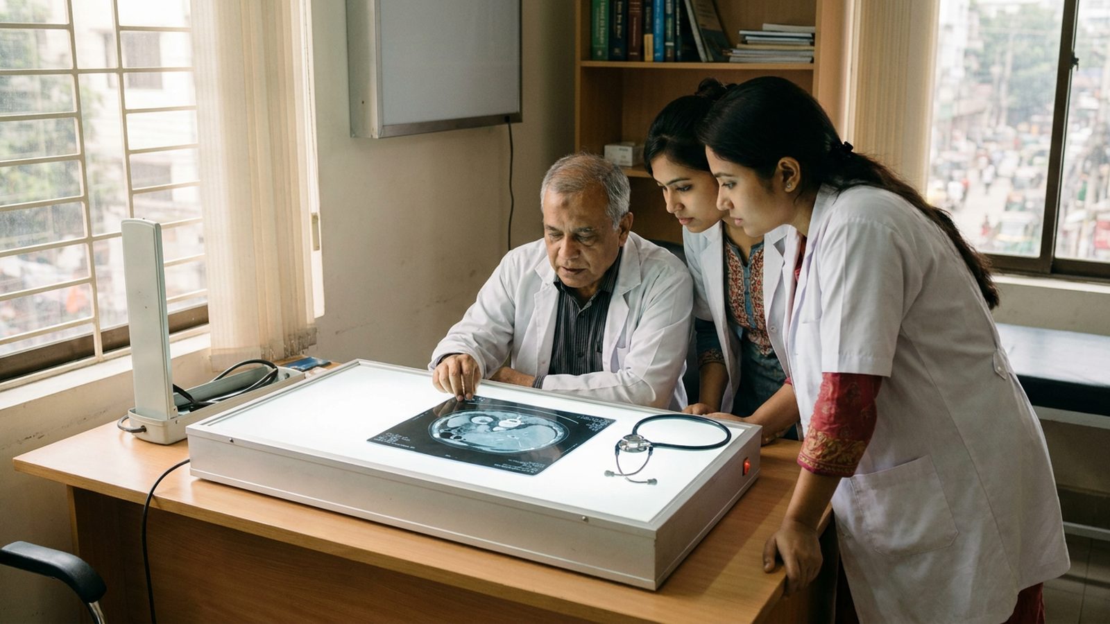
Bring the images, not only the reports. A CT or MRI on a disc or a shared link lets the vessels be traced and the remnant volume judged, which no typed summary can settle. Also bring any histopathology result, previous operation notes, a dated record of chemotherapy cycles, hepatitis serology with current treatment, and recent bilirubin, albumin, INR, platelets and creatinine.
Research-based practice: what Dr. Arefin has published
Dr. Murshidul Arefin is a research-active hepatobiliary surgeon, not only an operating one. He is a co-author of a peer-reviewed, open-access Cureus study on the preoperative albumin-bilirubin score as a predictor of complications after liver resection, which is the work quoted above, and of a case report on neuroendocrine tumour of the ampulla of Vater. That reading habit is why liver reserve is measured and stated plainly before a major resection is offered, rather than assumed from the size of the tumour.
Booking a Consultation or a Second Opinion
Consultations run at Popular Medical College Hospital in Dhanmondi, Dhaka. Call +880 1311 487 592 to Book an Appointment, or send the imaging and reports for review first if you are travelling in from outside the city. A second opinion is welcome, including cases where resection has been advised or refused elsewhere. If ablation, systemic treatment or transplant assessment is the better route, or no operation is appropriate, you will be told that plainly.
Frequently Asked Questions
How much liver can be safely removed?
It depends on the liver, not a fixed fraction. A healthy liver tolerates a much smaller remnant than a fibrotic, cholestatic or chemotherapy damaged one, which is why volumetry and function are assessed together.
Does the liver grow back after resection?
The remnant enlarges and takes over function over weeks, which is why the operation is possible at all. It does not regrow the original shape, and a cirrhotic liver regenerates poorly.
Can a major liver resection be done laparoscopically?
Selected resections can, depending on the tumour position and its relation to the vessels. Access is chosen for safety, and an open operation is not a lesser one.
Is surgery always better than ablation or chemotherapy?
No. Resection offers the best chance in selected patients, and ablation, transarterial therapy, systemic treatment or transplant assessment is more appropriate in others.
Written and reviewed by Dr. Murshidul Arefin, MBBS, FCPS, MS in Hepatobiliary Surgery (BSMMU), consultant at Popular Medical College Hospital, Dhanmondi, Dhaka, and co-author of peer-reviewed Cureus research on complications after liver resection.
Related Articles

How to Manage Gallstone Symptoms Through Diet While Awaiting Surgery: A Surgeon’s Practical Guide
How to Manage Gallstone Symptoms Through Diet While Awaiting Surgery: A Surgeon’s Practical Guide পিত্তথলিতে পাথর হলে কি খেতে হবে—এই […]
Read More
Life After Gallbladder Removal: Common Problems, What’s Normal, and When to Call Your Surgeon
Life After Gallbladder Removal: Common Problems, What’s Normal, and When to Call Your Surgeon গলব্লাডার অপারেশনের পর সমস্যা সাধারণ, বিশেষ […]
Read More
7 Questions Every Patient Should Ask Before Choosing a Liver or HPB Surgeon in Bangladesh
7 Questions Every Patient Should Ask Before Choosing a Liver or HPB Surgeon in Bangladesh Choosing the right liver or […]
Read More