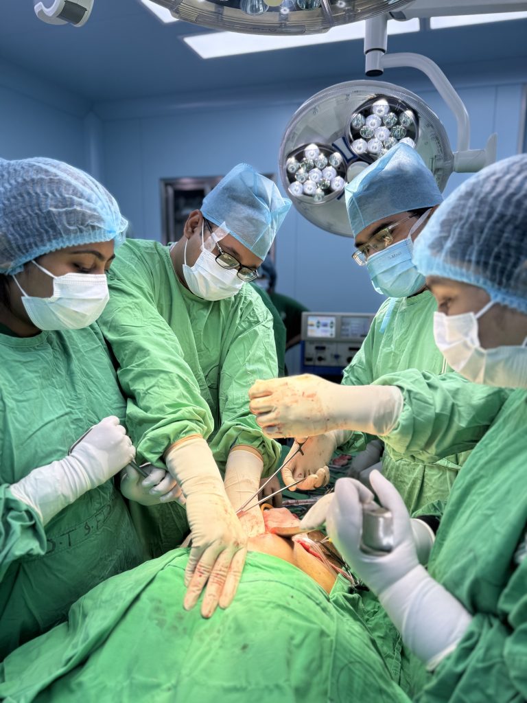
Biliary tract problems can present as intermittent pain, jaundice or life threatening infection, and early, appropriate treatment changes outcomes. This practical guide to biliary tract disorders treatment Dhaka walks patients through common symptoms and red flags, the diagnostic pathway from liver tests and ultrasound to MRCP and ERCP, and the endoscopic and surgical options available locally. You will also get clear advice on when to seek emergency care and how to choose a hepatobiliary surgeon and hospital in Dhaka, with practical steps to arrange evaluation at Popular Medical College Hospital with Dr Murshidul Arefin.
Overview of biliary tract disorders commonly seen in Dhaka
Straightforward fact: In Dhaka the bulk of referrals for biliary tract disorders treatment Dhaka are driven by complications of gallstones and by obstructive jaundice from stones or tumours. These conditions present on a spectrum from simple biliary colic to life threatening ascending cholangitis or locally advanced cholangiocarcinoma, and the correct pathway depends on the clinical urgency at presentation.
- Symptomatic gallstones: episodic right upper quadrant pain that leads patients to outpatient clinics.
- Acute calculous cholecystitis: inflamed gallbladder often requiring urgent laparoscopic cholecystectomy.
- Choledocholithiasis: stones in the common bile duct causing jaundice and biliary obstruction.
- Ascending cholangitis: fever, jaundice and hypotension needing immediate biliary drainage by ERCP or percutaneous means.
- Benign biliary strictures and post operative injuries: chronic cholestasis and recurrent cholangitis requiring reconstructive surgery.
- Cholangiocarcinoma and pancreatic head tumours: less common but high impact because delays convert resectable tumours into palliative cases.
Local context and practical considerations
Important consideration: Comorbid diabetes, delayed presentation and patchy access to advanced imaging change real outcomes in Dhaka. MRCP and ERCP exist in major centres, but patients referred from smaller hospitals often arrive after repeated symptomatic episodes or with infected bile, which increases perioperative risk and reduces options for single stage definitive care.
Trade off patients need to know: Endoscopic drainage by ERCP is fast and lifesaving in cholangitis, but it is not always curative for large impacted stones or complex hilar strictures where laparoscopic common bile duct exploration or formal biliary reconstruction may offer a more durable solution. Choose treatment based on what the centre can safely complete in one admission rather than on theoretical best case alone.
Concrete example: A 52 year old diabetic woman from Dhanmondi presented with fever, jaundice and severe right upper quadrant pain. Ultrasound showed dilated bile ducts and a suspected stone; she underwent emergency ERCP with stent placement to control infection and two weeks later had elective laparoscopic cholecystectomy and common bile duct clearance with complete recovery.
Early bile drainage in infection and timely staging for suspected cancer are the two single actions that most change outcomes for biliary disease in Dhaka.
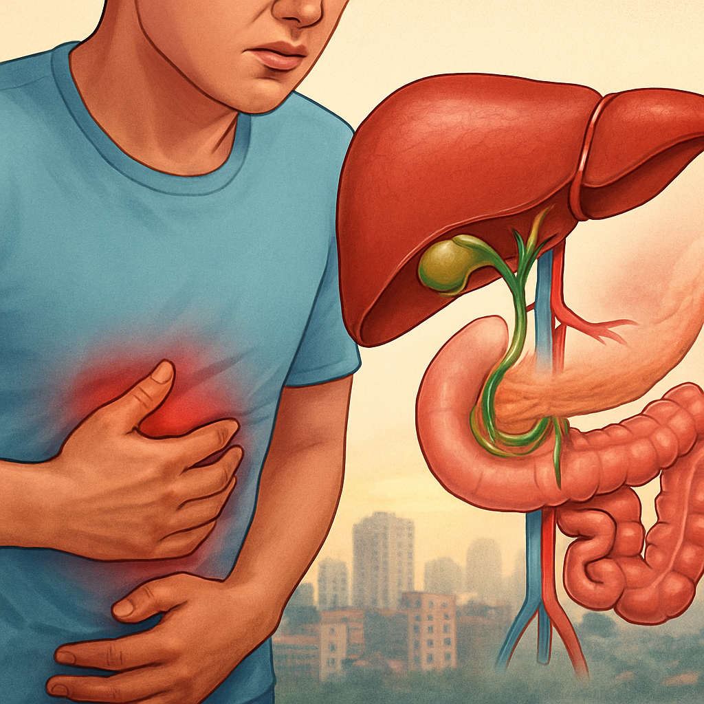
Clinical presentation and red flags that require urgent care
Immediate point: the clinical decision that matters is not the label but the physiology – whether the patient is septic, obstructed, or stable enough for imaging. Patients with systemic instability require action to decompress the biliary system and control infection before any definitive cancer staging or elective surgery.
Laboratory pattern to watch: a rising direct bilirubin with predominant alkaline phosphatase and GGT elevation indicates cholestatic obstruction. Marked transaminase elevation suggests another process such as viral hepatitis or hepatic ischemia and should prompt different urgent tests. Trends over 24 to 48 hours are informative – progressive bilirubin and worsening white cell count point toward failing drainage and need escalation.
Practical red flags and what to do first
- Fever plus jaundice and right upper quadrant pain: arrange immediate blood cultures, start broad spectrum IV antibiotics and plan for urgent biliary drainage – do not wait for MRCP if the patient is clinically deteriorating.
- Hypotension or altered mental status with jaundice: treat as severe sepsis; resuscitate, call the hepatobiliary team, and prepare for emergency ERCP or percutaneous transhepatic biliary drainage (PTBD) if ERCP is not immediately available.
- Rapidly rising bilirubin over days or new coagulopathy: this increases perioperative risk – obtain imaging and consult a hepatobiliary surgeon early to prioritise drainage and optimise liver function before any major operation.
Important limitation: abdominal ultrasound is a good first step but it misses distal common bile duct stones and small hilar strictures. In practice, a normal ultrasound does not rule out choledocholithiasis; MRCP or endoscopic ultrasound are often needed when clinical suspicion remains high.
Tradeoff to consider: if ERCP capability is limited after hours, PTBD provides reliable drainage but it carries different risks – external drains, catheter care and later conversion to internal stents or surgery. Choose the method a centre can complete safely that day rather than an ideal procedure that will be delayed.
Concrete example: a 68 year old man from Mirpur developed painless progressive jaundice and 6 kg weight loss over 8 weeks. He was icteric without fever. Rapid MRCP and contrast CT at the receiving hospital suggested a hilar mass; an early coordinated pathway arranged EUS guided biopsy and biliary stenting to improve liver function before multidisciplinary review for possible hepatectomy.
Judgment call clinicians make incorrectly: too often antibiotics are given and discharge planned without arranging definitive drainage or specialist review. In Dhaka settings, that delays care and raises risk of recurrent cholangitis. If there is any sign of systemic infection or worsening labs, escalate rather than observe.
If cholangitis is suspected, prioritise drainage over additional imaging – early ERCP or PTBD saves lives.
Diagnostic pathway and tests available at Popular Medical College Hospital
Direct statement: Popular Medical College Hospital runs a regimented, urgency‑based diagnostic pathway for biliary disease so decisions are made by physiology (obstructed, infected, stable) not by habit. Tests are staged to answer one question at a time: is there obstruction, does the patient need immediate drainage, and is there a resectable tumour?
Stepwise diagnostic workflow used in practice
- Rapid triage and basic labs: LFTs, CBC and inflammatory markers; blood cultures when febrile determine need for immediate drainage and antibiotic strategy.
- Point of care imaging: focused abdominal ultrasound by experienced sonographers to look for gallstones, ductal dilation or free fluid — quick but not definitive for distal or tiny stones.
- Cross sectional imaging: contrast CT abdomen when malignancy or vascular involvement is suspected; CT also helps plan major resections.
- Noninvasive duct imaging: MRCP for detailed biliary anatomy when time and renal function permit; useful for surgical planning and tumour mapping.
- Endoscopic assessment: EUS when ultrasound/MRCP are inconclusive for small stones or periampullary lesions; ERCP reserved where intervention (stone extraction, sphincterotomy, stent) is anticipated.
Practical tradeoff: MRCP is safe and avoids endoscopic risk but often requires scheduling and occasionally misses stones under a few millimetres. EUS is more sensitive for small choledocholithiasis and can be combined with ERCP in the same session — but only when interventional endoscopy and anaesthesia are reliably available that day.
Concrete example: A 45 year old man with intermittent painless jaundice had a normal abdominal ultrasound but cholestatic LFTs. He underwent EUS which found a 6 mm common bile duct stone; immediate ERCP with sphincterotomy and stone extraction was performed in the same admission and he went home the next day with normalising bilirubin.
Important: ERCP is therapeutic and commonly curative for obstructive cholangitis and choledocholithiasis, but it carries procedure‑specific risks; choose it when the expected benefit (rapid drainage or stone removal) outweighs those risks and local expertise exists.
Next consideration: When choosing a centre for biliary tract disorders treatment Dhaka, confirm 24/7 endoscopy coverage, on‑site MRCP/CT capability and access to an experienced HPB surgical team before you commit to definitive care.
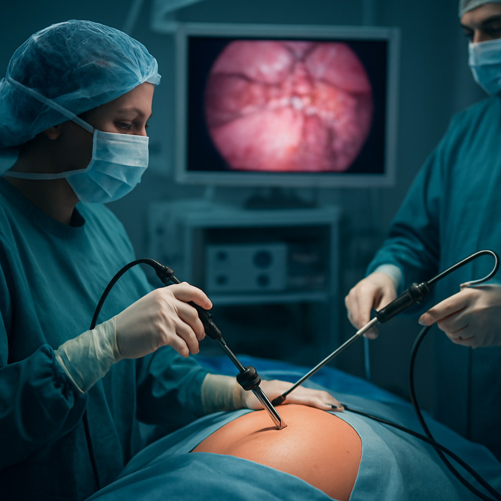
Surgical and endoscopic treatment options explained with indications
Immediate principle: choose the procedure that fixes the patient physiology you see now — urgent drainage for infection, targeted removal for obstructing stones, and planned resection for cancer. The technical best option matters less than whether the chosen centre can safely complete that step without repeated transfers or delays.
How to match procedure to problem in practice
| Procedure | Typical indication | Expected recovery / timeframe | Key limitation or tradeoff |
|---|---|---|---|
| ERCP with sphincterotomy ± stent | Ascending cholangitis, choledocholithiasis, temporary biliary decompression | Same‑day to 48 hours for uncomplicated cases | Risk of pancreatitis; may be incomplete for large impacted stones |
| Endoscopic ultrasound (EUS) guided interventions | Small distal stones not seen on MRCP, tissue diagnosis for periampullary lesions | Often same admission if combined with ERCP | Requires interventional endoscopy team and anaesthesia coordination |
| Laparoscopic cholecystectomy | Symptomatic gallstones, uncomplicated cholecystitis after optimization | Discharge typically 24–48 hours | Not a bile duct drainage procedure; concomitant CBD stones need separate management |
| Laparoscopic common bile duct (CBD) exploration | Definitive single‑stage clearance of CBD stones at time of cholecystectomy | Shorter total hospital pathway when feasible | Requires surgical expertise and equipment; longer operative time than standalone cholecystectomy |
| Roux en Y hepaticojejunostomy | Benign high strictures, major bile duct injury | Longer hospital stay and recovery over weeks | Major reconstruction with risk of anastomotic stricture long term |
| Hepatic resection / Pancreaticoduodenectomy | Resectable cholangiocarcinoma or pancreatic head cancer | Prolonged recovery; complex perioperative care | High resource requirement; needs multidisciplinary care and ICU support |
| Percutaneous transhepatic biliary drainage (PTBD) | When ERCP is impossible or fails, and urgent drainage is required | Immediate external drainage, later internalisation or surgery | External catheter care burden; risk of bleeding or infection |
Practical insight: in Dhaka settings the most common mistake is selecting an ideal procedure that the centre cannot finish promptly. For example, scheduling a laparoscopic bile duct exploration when there is no on‑site interventional endoscopy or ICU can convert a single problem into multiple admissions. Prioritise a plan that the hospital can execute in the index admission.
Concrete case: A 58 year old man presented with high fever and jaundice. Emergency ERCP achieved rapid biliary drainage and culture‑directed antibiotics controlled sepsis; two weeks later, after liver function recovery and staging, he underwent laparoscopic cholecystectomy with single‑session CBD exploration for definitive stone clearance and avoided a second hospital transfer.
Judgment that matters: ERCP is lifesaving for infected obstruction but it is not a substitute for oncologic resection when a tumour is the cause. Conversely, aggressive resections without prior optimisation or drainage in jaundiced febrile patients increase morbidity. Good outcomes come from sequencing interventions correctly — drain, stabilise, stage, then resect when appropriate.
If you need a rapid decision in Dhaka, confirm 24/7 ERCP capability and HPB surgical coverage at the same centre before committing to definitive surgery.
How to choose the right surgeon and hospital in Dhaka
Start with the problem you need solved, not the brand name. For biliary tract disorders treatment Dhaka the decisive factor is whether the centre can complete urgent drainage, diagnostic staging and the definitive procedure without risky transfers. A highly publicised surgeon is useful only if the hospital has on‑call interventional endoscopy, reliable imaging and postoperative critical care.
What to verify before you book
Key credentials to check: Look for a surgeon with advanced training in hepatobiliary and pancreatic surgery (fellowship or similar), regular case volume in HPB operations, and participation in multidisciplinary tumour boards. Experience matters most for complex operations such as hepatic resection or pancreaticoduodenectomy because perioperative judgement drives outcomes more than promotional language.
- Hospital capabilities: on‑call therapeutic endoscopy and anaesthesia, cross‑sectional imaging (contrast CT/MRCP) and interventional radiology available 24/7
- Support services: dedicated ICU beds, experienced anaesthesia team, and pathology/oncology input for cancer cases
- Team coordination: scheduled MDT meetings for suspected cholangiocarcinoma or complex strictures, and clear pathways for postop complications
Practical tradeoff: High‑volume tertiary units in Dhaka usually deliver better outcomes for major resections but may require longer scheduling and travel; smaller hospitals can be faster for straightforward ERCPs or laparoscopic cholecystectomy but may lack ICU or interventional radiology backup. Choose the option that matches the clinical urgency and the risk of transfer.
Concrete example: A 46 year old man with ascending cholangitis arrived at a neighbourhood clinic after hours. The clinic could not perform therapeutic endoscopy, and transfer to a tertiary hospital added three hours — during which his sepsis worsened. At an HPB centre with round‑the‑clock endoscopy and critical care the patient had prompt ERCP, stabilisation and same‑admission cholecystectomy once infection cleared.
Questions to ask on the phone: How many ERCPs and major HPB resections does the surgeon perform per year? Is there an on‑call interventional endoscopy service overnight? Will my case be reviewed in an MDT and is ICU required or available? Ask also about expected hospital stay and a realistic cost range for your specific procedure.
Local next step: For coordinated HPB care in Dhanmondi, you can review team services at Popular Medical College Hospital and arrange consultation via drarefin.com/appointment. Bring digital copies of prior scans and recent liver tests to make the first visit practical and efficient.
Priority judgement: prefer a centre that can safely complete the next necessary step that day — drain, stabilise or resect — rather than one that only offers an ideal procedure with likely delays.
Preoperative preparation and perioperative risk optimization
Core point: Proper preoperative optimisation prevents more complications than choosing between an open or laparoscopic approach. Focus on physiology first: correct infection, improve metabolic state, and confirm that the centre can deliver the planned intervention plus immediate rescue care.
Domains that change outcomes
Medical optimisation: Control hyperglycaemia, correct anaemia and manage fluid status. For patients with obstructive jaundice, improving cholestasis before a major resection lowers infectious and hepatic complications but delaying oncologic surgery has a tradeoff – the team must weigh short term perioperative risk against tumour progression.
- Preop labs and imaging current: recent LFTs, coagulation, cross sectional imaging within 4 weeks and documented bile drainage plan if bilirubin is rising
- Medication plan: anticoagulant/antiplatelet strategy agreed with anaesthesia; steroid or immunosuppressant adjustments noted
- Logistics reserved: ICU bed or high dependency unit on standby for major resections and availability of blood products and interventional radiology
- Nutrition and mobility: short course of prehabilitation for malnourished patients, and clear plans for early postoperative ambulation
Concrete example: A 60 year old man scheduled for right hepatectomy had bilirubin 6 mg/dL and poorly controlled diabetes. The team performed ERCP stent drainage, started a targeted glycaemic control plan and postponed major resection by three weeks. At operation his liver function had improved, wound infection risk fell and he left ICU earlier than expected.
Antibiotics and infected bile: Do not treat cultures as a substitute for drainage. In Dhaka practice we see resistant organisms from prior empirical treatment. Obtain bile and blood cultures at the time of drainage, start empiric IV antibiotics for cholangitis, then narrow therapy based on results to avoid selecting multidrug resistant bacteria.
Anaesthesia and hepatic reserve: Evaluate coagulation, synthetic function and cardiopulmonary fitness. For marginal liver reserve or extended resections consider portal vein embolisation or staged hepatectomy only at centres with hepatobiliary critical care. That judgement separates successful high risk cases from preventable failures.
Next step for patients in Dhaka: bring recent scans, LFTs and any bile culture reports to your HPB appointment. For coordinated preoperative planning and scheduling of ERCP, imaging and surgical dates use the HPB services link at Popular Medical College Hospital services or book via drarefin.com/appointment.
Postoperative recovery, follow up care and anonymized case summaries
Immediate assertion: Recovery after biliary procedures is defined by physiology and procedure type, not by a fixed day count. Patients who have ERCP recover differently and faster than those who have major liver or pancreatic resections, and your follow up plan should reflect that variation from day one.
Early recovery milestones and practical expectations
What to expect in the first 72 hours: After ERCP most patients stabilise within 24 to 48 hours unless there is post-ERCP pancreatitis or ongoing infection. After laparoscopic cholecystectomy expect pain that is controlled with oral analgesics, bowel function return within 24 to 48 hours and discharge the same or next day in uncomplicated cases. Major resections such as hepatic resection or pancreaticoduodenectomy require ICU monitoring, progressive mobilisation and a hospital stay measured in days to weeks.
Tradeoff to plan for: Early discharge reduces hospital acquired problems and cost, but premature outpatient care increases the chance of missed bile leak or sepsis. In Dhaka, where returning to hospital can be logistically hard for families, I err on the side of a longer in-hospital observation for high-risk patients and a clear direct line for rapid readmission if red flags appear.
Follow up schedule, surveillance and who does what
Typical follow up timeline: For routine ERCP or cholecystectomy a clinical review at 7 to 14 days and a blood test at 4 to 6 weeks is reasonable. For bile duct reconstruction or major oncologic resections plan early surgical clinic review at 7 to 10 days, repeat cross sectional imaging at 6 weeks to assess recovery, then oncology and imaging surveillance tailored by pathology results.
- Early tests: LFTs and CBC at first clinic visit to detect cholestasis or early infection
- Imaging: MRCP or contrast CT at 6 weeks for complex duct repairs or after cancer resection to document anatomy and early recurrence
- Long term surveillance: CT or MRCP frequency determined by final pathology; typical oncology follow up is every 3 to 6 months in the first two years
Important limitation: Access to MRCP or repeat CT in Dhaka can be delayed and costly. When advanced imaging is unavailable, clinical assessment and LFT trends guide early decisions—but never skip imaging when cancer recurrence is a concern. Use the hospital coordination service to time scans and clinic visits efficiently via drarefin.com/appointment.
Complication signals and immediate steps
Watch closely for these changes: increasing abdominal pain, fever, persistent vomiting, new or worsening jaundice, or bile draining from wounds. These are not minor; they require urgent reassessment and often early imaging and blood cultures. For suspected bile leak we perform ultrasound and contrast CT and consider ERCP for stent placement or percutaneous drainage depending on anatomy and stability.
Clinical judgement I use: Normalising symptoms with falling bilirubin is reassuring but not definitive for retained stones. If cholestatic LFTs plateau rather than fall, organise MRCP or EUS – retained stones are a common avoidable cause of readmission.
Anonymized case summaries from practice
Case A – ERCP for cholangitis: A middle aged woman presented febrile and jaundiced; emergency ERCP achieved biliary drainage and she received targeted antibiotics. She was discharged after 48 hours and returned two weeks later for elective laparoscopic cholecystectomy with no complications.
Case B – Laparoscopic cholecystectomy: A man with recurrent biliary colic had same admission laparoscopic cholecystectomy and went home the next day. Telephone follow up on day 3 identified a superficial wound seroma managed conservatively without readmission.
Case C – Pancreaticoduodenectomy: A patient with resectable pancreatic head cancer underwent Whipple procedure with planned ICU care. Early biliary stent placement before operation improved liver function; recovery took three weeks in hospital with staged chemotherapy planning and close nutrition support.
Next consideration: If you live outside central Dhaka, plan post discharge logistics in advance – transport, nearby lab access, and a named contact at the HPB clinic reduce the risk that a manageable complication becomes an emergency.
Related Articles
Laparoscopic Surgery: Benefits, Recovery and What Patients in Dhaka Should Expect
Laparoscopic Surgery: Benefits, Recovery and What Patients in Dhaka Should Expect In Dhaka, laparoscopic surgery often means smaller incisions, less […]
Read More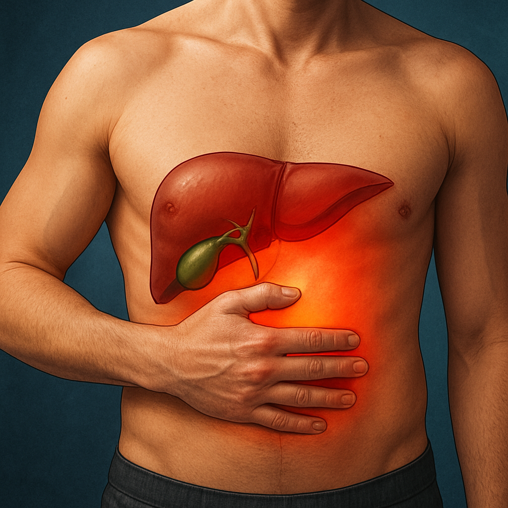
Gallbladder Problems: Signs, Treatment Options and When to See a Surgeon in Dhaka
Gallbladder Problems: Signs, Treatment Options and When to See a Surgeon in Dhaka In Dhaka, gall bladder problems can masquerade […]
Read More
Understanding Liver Function Tests: A Clear Guide for Patients in Dhaka
Understanding Liver Function Tests: A Clear Guide for Patients in Dhaka If you are in Dhaka and have jaundice, abdominal […]
Read More