Cholangiocarcinoma Surgery Dhaka for Bile Duct Cancer
When a scan report says there is a mass in the bile duct, the only question that matters that week is whether an operation is still possible, and that answer depends on where the tumour sits.
What Cholangiocarcinoma Is, and Where It Starts
Cholangiocarcinoma is a cancer that arises from the lining of the bile ducts, the thin channels that carry bile from the liver to the intestine. It is grouped by location, and that grouping decides almost everything about treatment. Intrahepatic tumours sit inside the liver itself. Perihilar tumours, often called Klatskin tumours, sit at the junction where the right and left ducts meet, which is the most technically demanding position of the three. Distal tumours sit in the lower part of the duct, close to the pancreas and the ampulla, and behave more like periampullary disease.
Moreover, location changes the operation completely. That is why cholangiocarcinoma surgery in Dhaka should never be planned from an ultrasound report alone. The tumour is uncommon compared with gallstone disease, but in a hepatobiliary practice it is not rare.
Key Takeaways for Cholangiocarcinoma Surgery:
- Painless yellowing of the eyes and skin in an adult needs urgent imaging, because a blocked duct from any cause does not improve by waiting.
- Tumour location, intrahepatic, perihilar or distal, decides the operation, so full duct mapping comes before any decision about surgery.
- A scan finding is not a diagnosis, since stones, strictures and inflammatory disease can produce the same blockage and the same jaundice.
- Some tumours are not removable at the time they are found, and identifying that on imaging is far better than discovering it during an operation.
- Deep jaundice is usually drained and nutrition corrected before major resection, because operating through it raises the risk of serious complications.
Painless Jaundice and Other Early Warning Signs
The classic presentation is yellowing of the eyes and skin that appears without pain. Patients notice dark urine, pale stools, and itching that is often worse at night. Weight loss and loss of appetite are common. Fever with chills means infection in a blocked duct, which is an emergency. In Bangla, families usually hear the phrase পিত্তনালীর ক্যান্সার, and the fear that follows it is entirely understandable.
However, the same picture can come from gallstones in the duct, from a benign stricture after previous surgery, from inflammatory duct disease, and from other causes that need surgery for a blocked bile duct rather than cancer treatment. Nobody should be told they have bile duct cancer on the strength of one ultrasound. If your reports already mention a duct mass or an unexplained block, Get a Second Opinion before any irreversible decision is taken, because the order of the next few tests genuinely changes what is possible later.
A Patient Experience in Dhaka
A man in his early sixties from Dhaka arrived with three weeks of painless jaundice and an outside plan to place a stent and review in two months.
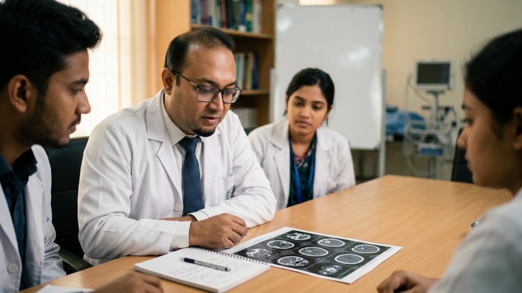
Rather than proceeding straight to a stent, the duct anatomy was mapped first with MRCP, then a contrast CT of the chest and abdomen was done to look at the vessels and check for spread. That sequence mattered: the imaging showed the tumour sat at the duct junction, and a stent placed carelessly at that level can make later reconstruction harder. Drainage was then done deliberately, on the side that would be kept, while nutrition and albumin were corrected over the following weeks. No promises were made to him about outcome, and none are made here. What he got was a plan built in the right order. Shared with permission, identifying details removed.
MRCP, CT and Staging Before Bile Duct Cancer Surgery
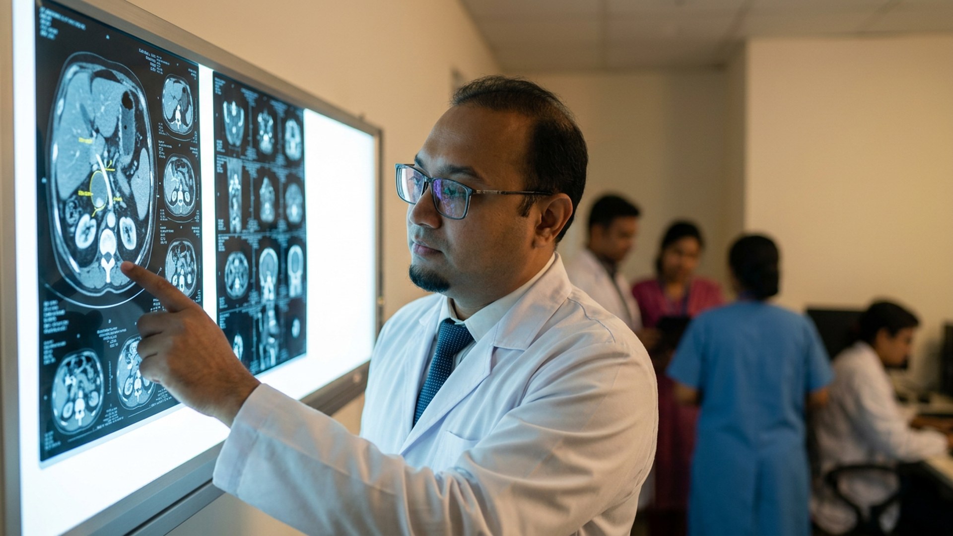
Ultrasound comes first and shows whether the ducts are dilated and where the block sits. MRCP then maps the biliary tree in detail, which is the single most useful study for deciding what operation is even on the table. A contrast CT of the chest, abdomen and pelvis follows, to assess involvement of the portal vein and hepatic artery and to look for disease outside the liver. Blood tests include bilirubin, albumin, full liver function and CA 19-9.
However, CA 19-9 deserves a warning. It rises in simple duct obstruction and in infection, so a high value in a jaundiced patient proves very little on its own and a normal value does not exclude cancer. Tissue confirmation is obtained where it can be taken safely, often by brushings at ERCP or by biopsy. General descriptions of the disease from the NHS and the general overview of cholangiocarcinoma are worth reading alongside your own reports.
Why Some Tumours Are Resectable and Others Are Not
Resectability is a judgement built from four things. First, how far the tumour spreads along the ducts. Second, whether the portal vein and hepatic artery are involved. Third, whether the part of the liver that would remain is big enough and healthy enough to carry the patient through recovery. Fourth, whether disease has already spread to distant sites or to the lining of the abdomen.
Furthermore, this is where honesty matters more than optimism. A meaningful proportion of bile duct cancers are already beyond removal when they are first found, and detecting that on good imaging is far better than opening an abdomen to discover it. Anybody offering bile duct cancer surgery Bangladesh wide without full vascular imaging and a remnant liver assessment is not planning, they are gambling. In control of that decision should be a full set of images reviewed properly, alongside the patient and family who deserve to hear the reasoning.
Resection and Reconstruction Options by Tumour Location
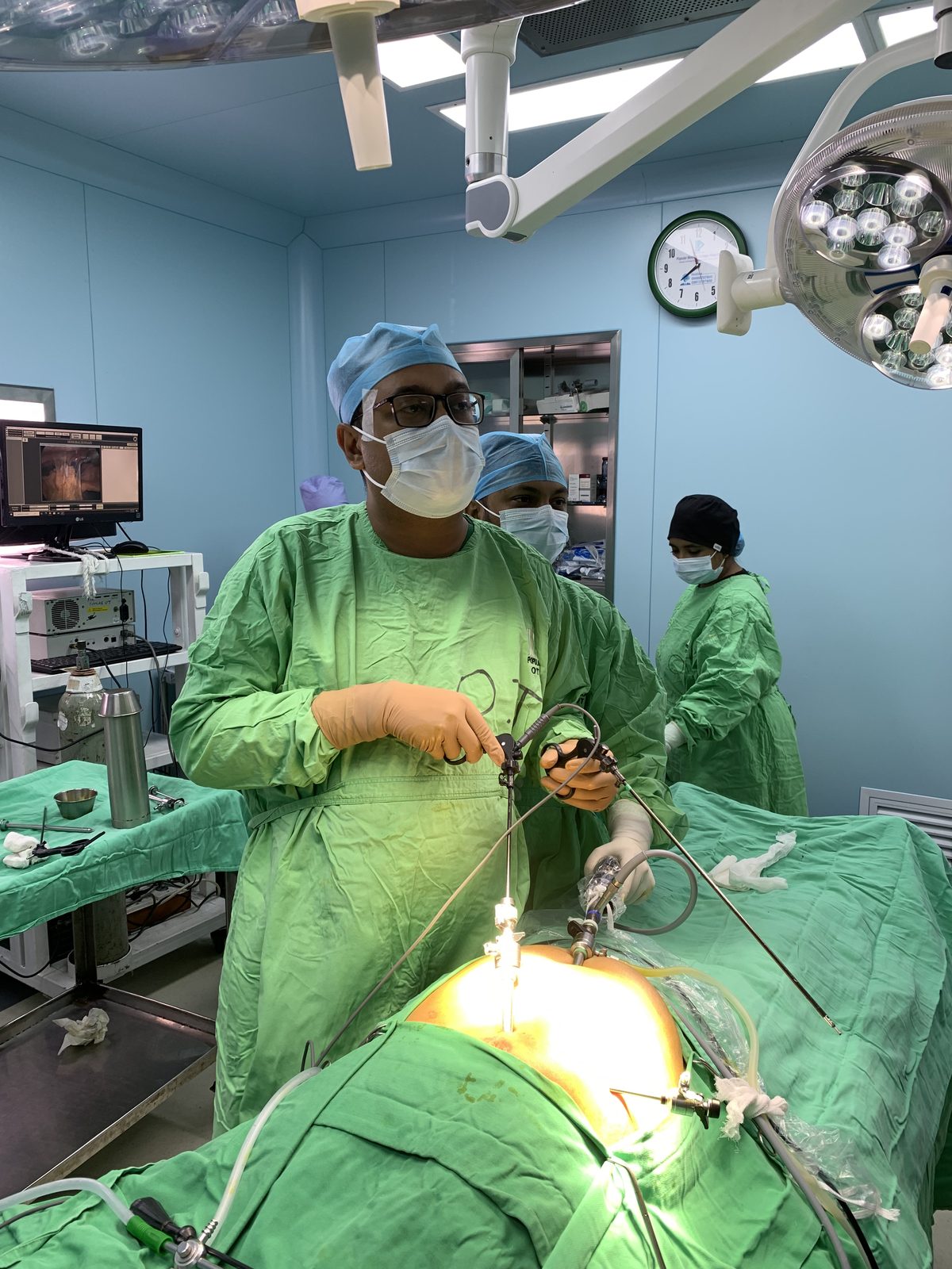
For distal tumours in the lower duct, the standard operation is a Whipple procedure, removing the head of the pancreas, the lower bile duct and the duodenum, then reconstructing to the small bowel. For perihilar tumours, the operation is bile duct excision combined with liver resection on the affected side, with reconstruction by hepaticojejunostomy, joining the remaining healthy duct to a loop of jejunum. For intrahepatic tumours, the work is liver resection aiming for clear margins, with the extent decided by tumour position and remnant volume.
Moreover, lymph node clearance is part of all three, since node status drives what oncology recommends afterwards. Reconstruction is unforgiving work: the duct is thin and a join under tension leaks or narrows later. The practice behind this page rests on 200+ bile duct reconstructions with an 85% referral rate and 300+ complex pancreatic and upper-GI cases with a greater than 95% success rate. Those figures describe experience in similar bile duct surgery, not a prediction about any individual result.
Drainage, Nutrition and Preparation Before Surgery
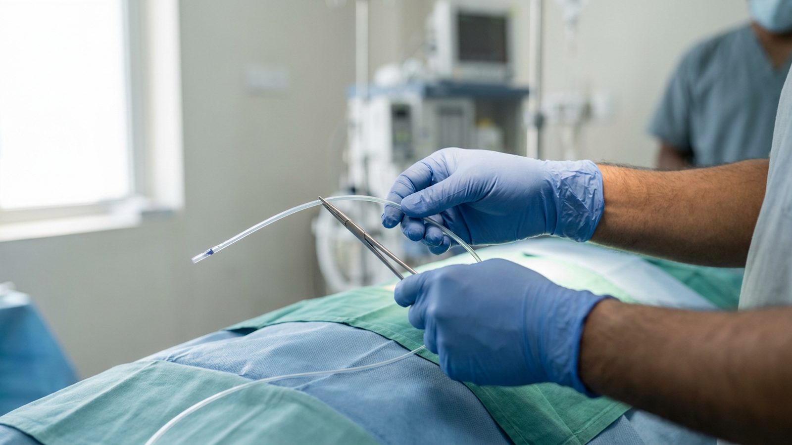
Major resection through deep jaundice is a poor idea, and the reason is simple: a liver soaked in bile heals badly, clots badly and tolerates resection badly. So when bilirubin is high and a big operation is planned, the duct is drained first, either endoscopically or through the skin, and drainage is directed at the side of the liver that is going to be kept.
Furthermore, the weeks between drainage and surgery are working weeks, not waiting weeks. Albumin and nutrition are corrected, often with high protein intake and supplements, because a patient who has lost eight kilograms will not recover from a Whipple on willpower. Infection is controlled properly before any incision. In addition to all of that, existing conditions such as diabetes and heart disease are stabilised, which is usually where a delay of two weeks buys far more safety than it costs.
Peer-Reviewed Periampullary and Liver-Resection Research
Two published papers are directly relevant here, and both are co-authored rather than led, which is worth stating plainly.
The first is a co-authored Cureus case report on a neuroendocrine tumour of the ampulla of Vater, published in 2023. It describes surgery for a tumour in the periampullary region, the same anatomical territory as a distal bile duct cancer. A case report is one patient, so it demonstrates familiarity with the anatomy and the operation, not superiority of any technique, and it should not be read as anything more.
The second is a co-authored Cureus study on the ALBI score and complications after liver resection. The reported area under the curve was 0.659, which is weak to moderate discrimination and is described that way in the paper. Post-hepatectomy liver failure occurred in 0% of ALBI-1 patients, 38.5% of ALBI-2 patients and 100% of ALBI-3 patients in that series. However, the practical value is in planning rather than prediction: perihilar and intrahepatic resections take part of the liver away, so a preoperative marker of poor liver reserve is a reason to prepare harder or to reconsider the extent of resection, not a reason to refuse anybody an assessment.
Recovery, Nutrition and Oncology Follow-Up
Recovery from major hepatobiliary resection is measured in weeks, and honest expectation setting prevents a lot of distress. Expect drains for a period, gradual reintroduction of food, and walking on the first days rather than bed rest. A Whipple or a major liver resection means an extended hospital stay and weeks of low energy at home.
In addition, the operation is one part of treatment rather than the whole of it. Final histology, including margins and node status, is reviewed and then discussed with medical oncology, since adjuvant chemotherapy decisions rest on that report. Surveillance imaging and blood tests continue long term. Call immediately after discharge for fever, worsening or returning jaundice, severe abdominal pain, bile leaking from a wound or drain, or persistent vomiting. None of this is a promise about cure, and any surgeon who offers you one on a first visit is not being straight with you.
“Cholangiocarcinoma, also known as bile duct cancer, is a type of cancer that forms in the bile ducts.”
Source: Cholangiocarcinoma, Wikipedia
When Surgery Is Not the Answer
Sometimes the honest conclusion after full imaging is that removal is not possible, and saying so is part of the job. In that situation the aim shifts to relieving the block and keeping you comfortable and fed. Stenting, endoscopically or through the skin, drains bile so that jaundice, itching and appetite improve. Medical oncology then advises on chemotherapy and, in selected cases, radiotherapy or targeted treatment based on histology.
However, none of that is abandonment. Avoiding an operation that cannot remove the disease is a decision taken in your favour, because a futile laparotomy costs weeks of strength and returns nothing. The family is told the reasoning in plain language, with the imaging on the screen, so that decisions about travel abroad or further opinions can be made on facts rather than fear. Society guidance from SAGES and liver-disease guidance from EASL describe the same reasoning in professional terms.
Research-based practice: what Dr. Arefin has published
Dr. Murshidul Arefin is a research-active hepatobiliary surgeon, not only an operating one. He is a co-author of peer-reviewed, open-access studies in Cureus on albumin-bilirubin risk scoring before liver resection, alongside a case report on neuroendocrine tumour of the ampulla of Vater. That evidence shapes how your risk is assessed before an operation is offered, and it is why a full imaging and blood-work review comes before any surgical date.
Sending Your Reports for an Opinion in Dhanmondi
If you want cholangiocarcinoma treatment Dhaka options assessed properly, send the images rather than only the reports. MRCP and contrast CT studies on a disc or a shared link are what allow the duct anatomy and the vessels to be judged. Include recent blood results with bilirubin, albumin, liver function and clotting, any biopsy or brushing report, a list of current medicines, and a short note of what has already been done.
Furthermore, consultations run at Popular Medical College Hospital in Dhanmondi, Dhaka. Call +880 1311 487 592 to Book an Appointment, or send your reports for a second opinion before travelling if you live outside the city. If the answer after review is that surgery is not advisable, you will be told that directly, with the reason.
Frequently Asked Questions
Is bile duct cancer always inoperable?
No. Operability depends on tumour location, vessel involvement, remnant liver volume and spread. Some cases are removable, some are not, and only full imaging can separate the two honestly.
Should a stent be placed straight away?
Not always, and not blindly. Duct anatomy is mapped first when surgery may be possible, because a stent placed at the wrong level can complicate later reconstruction. Infection or very deep jaundice does change that order.
What operation would I need?
Distal tumours usually need a Whipple procedure, perihilar tumours need duct excision with liver resection and hepaticojejunostomy, and intrahepatic tumours need liver resection.
How long is recovery after surgery?
Weeks rather than days, with an extended hospital stay and a gradual return of appetite and energy at home. Histology review and an oncology discussion follow the operation.
Reviewed by Dr. Murshidul Arefin, MBBS, FCPS, MS in Hepatobiliary Surgery (BSMMU), hepatobiliary consultant at Popular Medical College Hospital, Dhanmondi, Dhaka, and a research-active surgeon published in Cureus.
Related Articles

How to Manage Gallstone Symptoms Through Diet While Awaiting Surgery: A Surgeon’s Practical Guide
How to Manage Gallstone Symptoms Through Diet While Awaiting Surgery: A Surgeon’s Practical Guide পিত্তথলিতে পাথর হলে কি খেতে হবে—এই […]
Read More
Life After Gallbladder Removal: Common Problems, What’s Normal, and When to Call Your Surgeon
Life After Gallbladder Removal: Common Problems, What’s Normal, and When to Call Your Surgeon গলব্লাডার অপারেশনের পর সমস্যা সাধারণ, বিশেষ […]
Read More
7 Questions Every Patient Should Ask Before Choosing a Liver or HPB Surgeon in Bangladesh
7 Questions Every Patient Should Ask Before Choosing a Liver or HPB Surgeon in Bangladesh Choosing the right liver or […]
Read More