
Choledochal Cyst Surgery Dhaka | Dr. M. Arefin

Choledochal Cyst Surgery Dhaka | Dr. M. Arefin

Choledochal Cyst Surgery Dhaka: Excision and Repair

The honest answer to “does this cyst really have to come out?” is usually yes, and the reason has less to do with today’s pain than with what the duct lining does over the next twenty years.

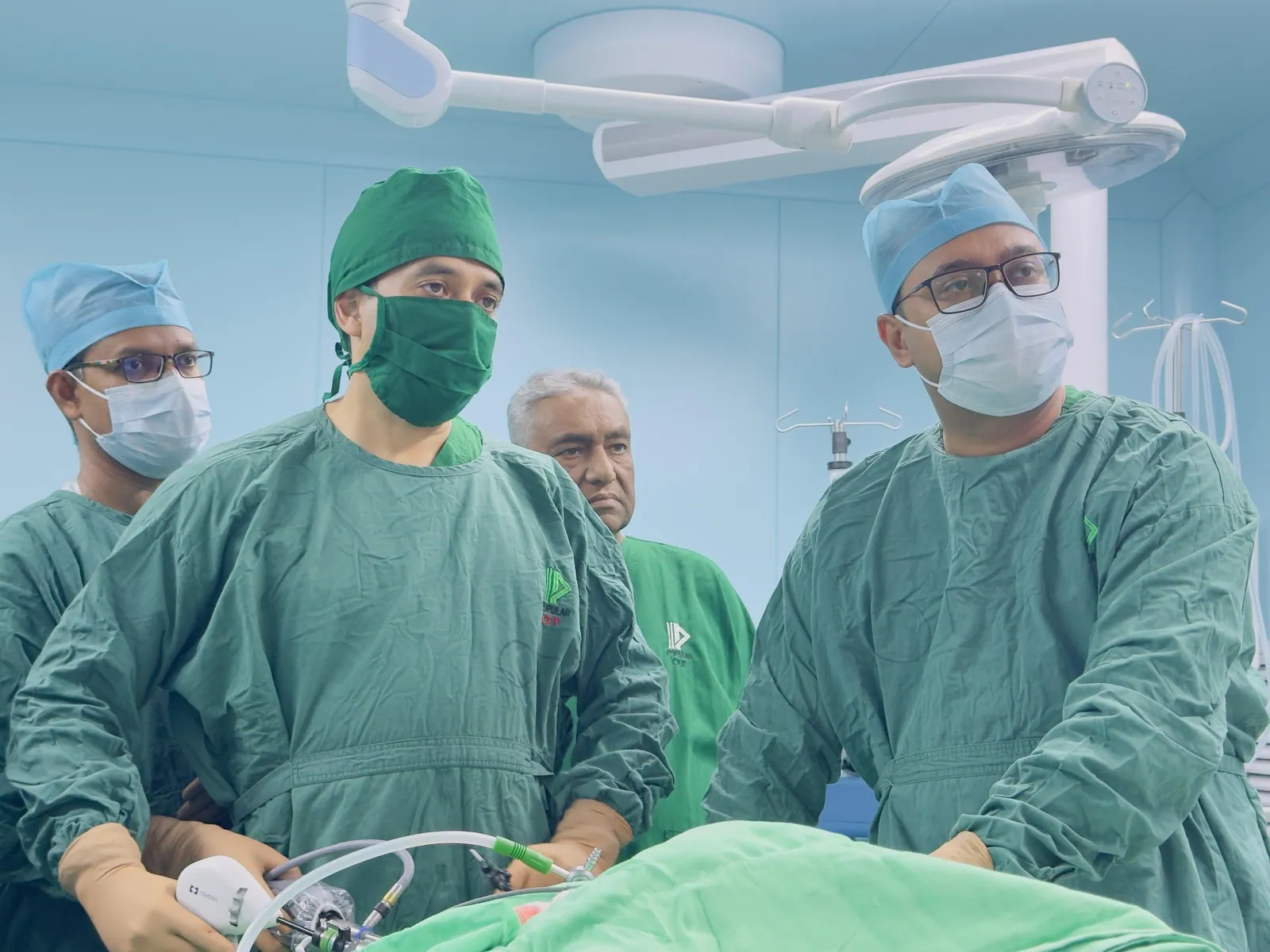
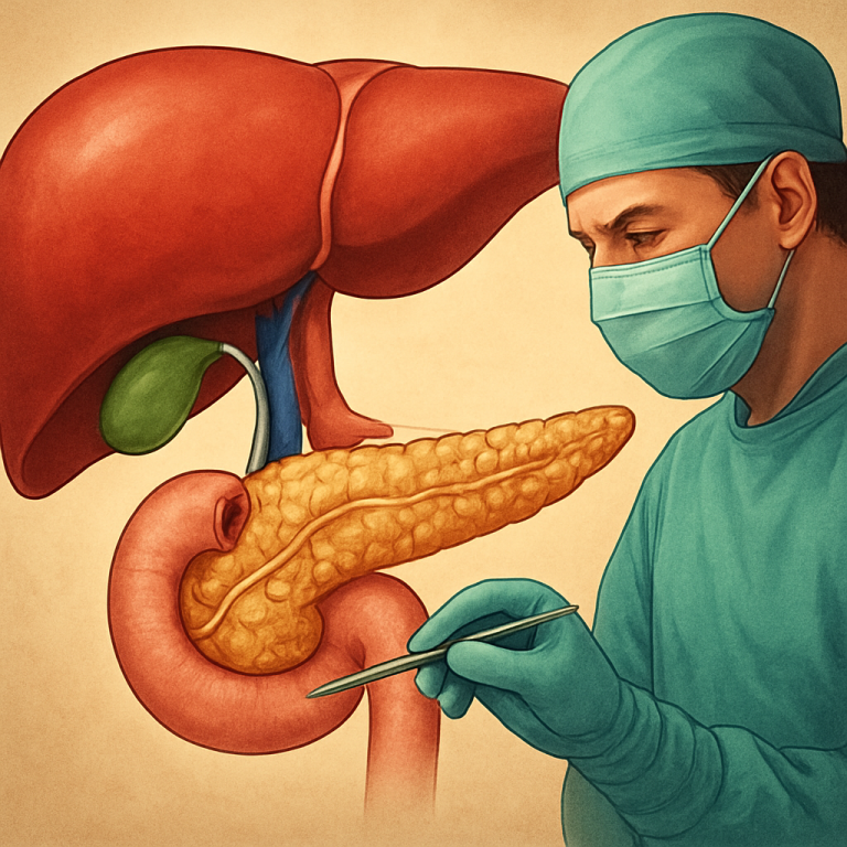
Clinicians performing laparoscopic choledochal cyst surgery Dhaka in a sterile operating theatre.
Dr. Murshidul Arefin and his surgical team perform a minimally invasive procedure using advanced laparoscopic instruments.

What a Choledochal Cyst Is, and Why It Is Treated

A choledochal cyst is a dilatation of the bile duct that a person is born with. The duct that should be a narrow channel carrying bile from the liver to the intestine is ballooned instead, along its whole length or in a segment. Many are found in childhood after repeated pain or jaundice, but a good number are diagnosed first in adults treated for years for something else.

The underlying problem is usually the junction where the bile duct and pancreatic duct meet. When it sits abnormally, pancreatic juice refluxes into the bile duct and bile flows into the pancreatic duct. Over years that reflux inflames the duct wall, which is why these cysts cause repeated cholangitis, pancreatitis and stones rather than sitting quietly. Moreover, chronic inflammation of the lining carries an increased long term risk of cancer in the wall itself, the main reason surgeons who work in biliary tract surgery recommend removal rather than observation.

Key Takeaways for Choledochal Cyst Surgery:

  • A choledochal cyst is a congenital dilatation of the bile duct, not diet related, and it does not resolve on its own.
  • Abnormal drainage where the bile and pancreatic ducts meet drives reflux, inflammation, stones and repeated attacks.
  • Treatment is advised because of repeated infection, pancreatitis and an increased long term cancer risk in the lining, not symptoms alone.
  • Complete removal with a new duct to bowel join is the accepted operation, since drainage leaves the abnormal lining behind.
  • Fever with jaundice is an emergency, so go to hospital rather than waiting for a clinic slot.
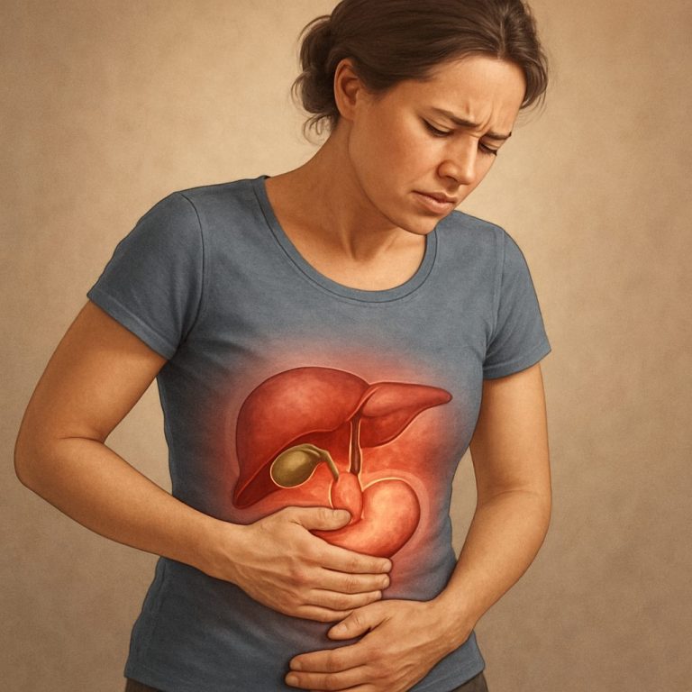
A South Asian patient discusses abdominal symptoms related to choledochal cyst surgery Dhaka with a specialist in a ward.
Early diagnosis of biliary symptoms is critical for successful choledochal cyst management in both children and adults.

Pain, Jaundice and a Lump: How It Presents

The textbook triad of upper abdominal pain, jaundice and a lump under the right ribs is complete in only a minority. In infants it is jaundice that persists or returns, pale stools and dark urine, sometimes with poor weight gain. In older children and adults the usual story is recurrent right upper abdominal pain over months or years, with occasional yellowing of the eyes, nausea and vomiting, and sometimes pancreatitis. Families in Bangladesh often hear the term পিত্তনালীর সিস্ট after the first ultrasound, usually without much explanation of what follows.

However, the commonest thing is delay. Episodic upper abdominal pain gets labelled gastric trouble and treated with acid suppression for years, and an ultrasound is requested only when jaundice appears or an attack lands the patient in hospital. Fever with chills alongside jaundice means infection in an obstructed duct and needs emergency assessment, not a clinic date. If a scan report already mentions a dilated duct, a cystic dilatation, or a blocked bile duct without a clear cause, Book an Appointment and bring the images themselves rather than just the typed reports.

A Patient’s Experience in Dhaka

A woman in her mid twenties from Dhaka was treated for gastritis for almost four years.

Her pain came in bouts of a few days, occasionally with mild yellowing the family put down to weakness. An ultrasound during one attack showed a dilated bile duct, and MRCP showed a fusiform cyst of the extrahepatic duct with an abnormal junction below it. She had complete cyst excision with a Roux loop hepaticojejunostomy, and the specimen went for full histology as it always should. The point is not the operation, which was standard, but the four years of acid tablets before it. Shared with permission, identifying details removed.

Ultrasound, MRCP and Cyst Classification

Ultrasound is the first test and usually raises the question, showing a dilated duct or a cystic space at the porta. It cannot, however, map the anatomy well enough to plan an operation. MRCP is the study that matters. It shows the shape and extent of the dilatation, whether intrahepatic ducts are involved, whether stones sit inside, and in many cases the abnormal junction itself. A contrast CT is added when a complication or a wall mass is suspected, or when vessels need checking before a difficult dissection. Blood tests include bilirubin, liver function, amylase or lipase if pancreatitis is in the picture, and a blood count when infection is suspected.

Furthermore, the shape of the cyst decides the operation, so classification is not academic. The Todani system gives five types. Type I, a fusiform or cystic dilatation of the extrahepatic duct, is by far the commonest. Type II is a diverticulum off the duct. Type III, a choledochocele, sits inside the duodenal wall. Type IV involves both extrahepatic and intrahepatic ducts. Type V, Caroli disease, is intrahepatic and a different planning problem, sometimes needing liver resection rather than duct excision alone. General descriptions from the NHS and an encyclopaedic overview of choledochal cysts are worth reading alongside your own reports.

Why Complete Excision Beats Drainage or Bypass

Older operations drained the cyst into the duodenum or a loop of bowel and left it in place. They relieved symptoms for a while, which is why they were popular, and they are now avoided by anybody who follows the disease long term, because the abnormal lining stays behind. Reflux continues, stones form in the retained cyst, cholangitis returns, and the cancer risk in that lining is not removed by improving bile flow past it.

Accepted practice for choledochal cyst excision Bangladesh wide, and internationally, is complete removal of the extrahepatic cyst with a new join between healthy duct and bowel. However, patients who had a drainage operation years ago are not rare, and revision surgery is harder: planes are scarred, anatomy is distorted, and the duct available for reconstruction is often short and inflamed. The retained specimen also has to be assessed for malignancy, so the operation is diagnostic as well as reconstructive. Anyone offered a quick internal drainage today should ask why the cyst is not being removed.

What the Operation Involves, Step by Step

Choledochal cyst surgery in Dhaka follows the same sequence as anywhere else. The cyst is exposed and dissected carefully off the portal vein and hepatic artery, which lie immediately behind it and are why this is not routine general surgery. Because the wall is inflamed the plane can be adherent, and in difficult cases the lining is stripped rather than risking the vein. The duct is divided above the cyst in healthy tissue near the hilum, so no abnormal lining is left behind. Below, the duct is divided close to the pancreas, without injuring the pancreatic duct or leaving a long stump that can later fill with debris.

Reconstruction is a Roux loop hepaticojejunostomy: a limb of jejunum joined to the healthy duct, single layer, tension free, with the blood supply of the duct end respected. In selected patients with favourable anatomy the dissection can be laparoscopic, which suits smaller uninflamed cysts better than scarred ones. The experience behind this page includes 200+ bile duct reconstructions with an 85% referral rate, alongside 800+ laparoscopic cases within over 1,500 surgeries. Those numbers describe the volume of similar bile duct surgery, not a prediction about your result.

Recovery in Children and Adults

Recovery after bile duct cyst surgery Dhaka families are quoted is measured in weeks, not days. Expect a hospital stay of several days, a drain near the new join, and feeding restarted gradually once the bowel wakes up. Walking starts on the first day or two, because lying still causes chest problems and leg clots rather than protecting the repair.

Children generally recover faster than adults, and that is not just youth. A child operated on before years of repeated cholangitis has cleaner tissue, a healthier duct and less scarring, so the join heals in better conditions. Adults with a decade of attacks, or a previous drainage operation, take longer. School or work usually resumes within a few weeks for uncomplicated cases, with heavy lifting avoided for longer. Diet needs no exotic restriction, and any fever, worsening pain, vomiting or returning jaundice at home is a reason to call rather than wait.

A Research-Active Hepatobiliary Practice in Dhaka

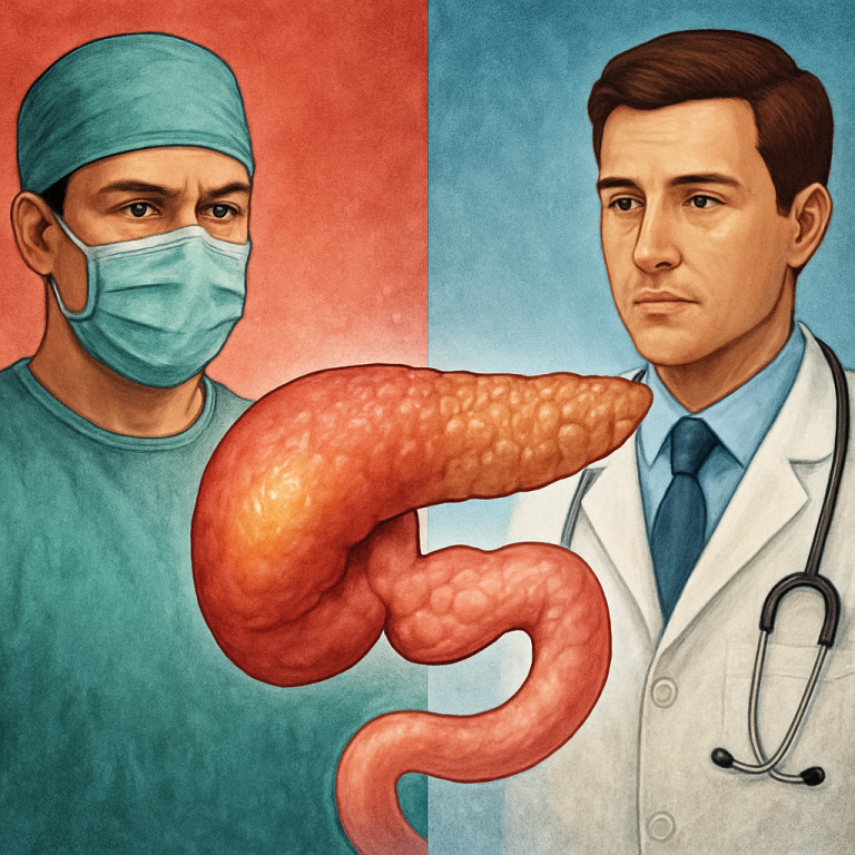
Rare conditions are where a reading habit shows. Choledochal cysts are uncommon enough that no surgeon accumulates a large personal series quickly, so judgement comes partly from published work rather than memory.

One directly relevant publication is a co-authored Cureus case report on a neuroendocrine tumour of the ampulla of Vater, published in 2023. Dr. Arefin is a co-author on that paper rather than the lead author, and it is described that way. Its relevance is the decision making it documents for a rare lesion in the periampullary region, the same territory as the lower end of a choledochal cyst. However, a case report is a single patient. It shows familiarity with the anatomy, imaging and operation, and that the work was reviewed by peers. It does not show that any technique is superior. Three peer reviewed Cureus papers sit behind this practice in total.

Long Term Follow Up and What Still Needs Watching

Removal of the cyst is not the end of the story, and any surgeon who discharges you permanently at six weeks is doing you a disservice. A hepaticojejunostomy can narrow over time, and a narrowed join causes recurrent cholangitis: episodes of fever, pain and jaundice months or years later. Stones can form above a tight anastomosis or inside intrahepatic ducts that were already abnormal, particularly in type IV and V disease. If lining was retained at a previous drainage operation, the cancer risk there remains and needs surveillance.

Controlling that risk needs straightforward follow up rather than anything elaborate. Reviews cover symptoms, then liver function tests, then an ultrasound, with MRCP when results or symptoms suggest a problem. Patients are also told exactly which symptoms bring them back early, since a fever with jaundice five years after a good operation still means an obstructed system until proven otherwise. Society resources from SAGES and liver-disease guidance from EASL describe the same follow up logic.

“A choledochal cyst is a congenital condition involving cystic dilatation of bile ducts.”

Source: Choledochal cysts, Wikipedia

Risks, Complications and Honest Limits

This is a real operation on a fragile structure, and risks are discussed before consent, not after a problem. Bile can leak from the new join early, usually managed with the drain left for exactly that reason. The join can narrow later, causing cholangitis. Bleeding is possible because the cyst sits on the portal vein and hepatic artery. The pancreas can be injured during division of the lower duct. Wound infection, chest infection and adhesive bowel obstruction years later all belong on the list.

However, none of that argues for leaving a symptomatic cyst alone, because the risks of repeated cholangitis, pancreatitis and retained abnormal lining accumulate year after year. What can honestly be offered is careful case selection, correct timing, unhurried dissection and long follow up, not a promise that nothing will go wrong.

Research-based practice: what Dr. Arefin has published

Dr. Murshidul Arefin is a research-active hepatobiliary surgeon, not only an operating one. He is a co-author of a peer-reviewed, open-access case report in Cureus on neuroendocrine tumour of the ampulla of Vater, work in the same periampullary territory as the lower end of a choledochal cyst. That reading habit shapes how your imaging and blood work are assessed before an operation is offered, and it is why the duct anatomy is mapped fully before any surgical date.

How to Book a Choledochal Cyst Consultation

Bring the images, not only the reports. Ultrasound and MRCP studies on a disc or shared link let the duct anatomy and the junction be judged properly, and a typed summary rarely settles the cyst type. Include recent blood results, previous operation notes, records of stents or drainage, current medicines, and for a child the weight record.

Consultations run at Popular Medical College Hospital in Dhanmondi, Dhaka. Call +880 1311 487 592 to Book an Appointment, or send the reports for review first if you are travelling in from outside the city. If surgery is not needed yet, or a different problem explains the pain, you will be told that plainly. A second opinion is welcome before any decision.

Frequently Asked Questions

Can a choledochal cyst be left alone?

Rarely, and only after careful assessment. Repeated infection, stone formation, pancreatitis and the long term cancer risk in the lining are why removal is usually advised rather than observation.

Is drainage of the cyst an alternative to removing it?

Not in current practice. Drainage leaves the abnormal lining in place, so reflux, stones, cholangitis and cancer risk continue. Complete excision with a duct to bowel join is accepted.

Can it be done laparoscopically?

In selected cases with favourable anatomy, yes. Heavily inflamed or previously operated cysts are usually safer open, and that decision is made on the imaging and at surgery.

How long is follow up after surgery?

Long term. The new join can narrow years later, so symptom review, liver function tests and imaging when indicated continue past six weeks.

Written and reviewed by Dr. Murshidul Arefin, MBBS, FCPS, MS in Hepatobiliary Surgery (BSMMU), consultant at Popular Medical College Hospital, Dhanmondi, Dhaka, and a research-active surgeon published in Cureus.

Scroll to Top