
Gallbladder infection, medically known as cholecystitis, is a condition characterized by inflammation of the gallbladder, often due to the presence of gallstones. This organ, located beneath the liver, plays a crucial role in the digestive process by storing bile, a substance that aids in the digestion of fats. When gallstones obstruct the bile ducts, they can lead to a buildup of bile within the gallbladder, resulting in inflammation and infection. The condition can manifest acutely or chronically, with acute cholecystitis being a medical emergency that requires prompt attention.
The prevalence of gallbladder infections varies across different populations, influenced by factors such as diet, genetics, and lifestyle. While some individuals may experience mild symptoms that resolve on their own, others may face severe complications if left untreated. Understanding the underlying mechanisms, risk factors, and treatment options for gallbladder infections is essential for both healthcare providers and patients alike. This article aims to provide a comprehensive overview of gallbladder infections, from clinical evaluation to prevention strategies.
In the context of understanding gallbladder infections, it is essential to explore various management strategies and clinical evaluations. A related article that delves into effective approaches for addressing gallbladder-related issues can be found here: Crafting Captivating Headlines. This resource provides insights that complement the discussion on imaging algorithms and antibiotic-to-surgery management strategies, enhancing the overall comprehension of gallbladder infection treatment.
Clinical Evaluation of Gallbladder Infection
The clinical evaluation of a suspected gallbladder infection begins with a thorough patient history and physical examination. Healthcare providers typically inquire about the patient’s symptoms, including abdominal pain, nausea, vomiting, and changes in appetite. The location and intensity of the pain are particularly important; patients often describe the pain as being located in the upper right quadrant of the abdomen and may report that it worsens after eating fatty foods. A physical examination may reveal tenderness in the same area, and signs such as Murphy’s sign—wherein the patient experiences pain upon deep inspiration while the examiner palpates the gallbladder—can further indicate cholecystitis.
Laboratory tests are also integral to the clinical evaluation process. Blood tests may reveal elevated white blood cell counts, indicating an inflammatory response, as well as liver function tests that can suggest bile duct obstruction or liver involvement. Additionally, imaging studies play a pivotal role in confirming the diagnosis. Ultrasound is often the first-line imaging modality due to its non-invasive nature and ability to visualize gallstones and assess gallbladder wall thickness. In some cases, computed tomography (CT) scans or magnetic resonance imaging (MRI) may be employed for further evaluation.
Imaging Algorithm for Gallbladder Infection Diagnosis

The imaging algorithm for diagnosing gallbladder infection typically begins with an abdominal ultrasound, which is considered the gold standard for initial assessment. This modality allows for the visualization of gallstones, thickening of the gallbladder wall, and fluid accumulation around the gallbladder—all indicative of cholecystitis. If ultrasound findings are inconclusive or if there is a suspicion of complications such as perforation or abscess formation, further imaging may be warranted.
In cases where ultrasound results are ambiguous or when there is a need for more detailed anatomical information, a CT scan can provide a comprehensive view of the abdominal organs. CT imaging is particularly useful in identifying complications associated with gallbladder infections, such as pancreatitis or biliary obstruction. Magnetic resonance cholangiopancreatography (MRCP) may also be utilized to visualize the biliary tree and assess for any blockages that could be contributing to the infection. By following this structured imaging algorithm, healthcare providers can accurately diagnose gallbladder infections and determine appropriate management strategies.
Antibiotic-to-Surgery Management Strategy for Gallbladder Infection

The management of gallbladder infections often involves a combination of antibiotic therapy and surgical intervention. Initial treatment typically begins with broad-spectrum intravenous antibiotics to combat infection and prevent further complications. The choice of antibiotics may be guided by local resistance patterns and individual patient factors. In many cases, empirical therapy is initiated while awaiting culture results to tailor treatment more specifically.
Surgical intervention is frequently indicated in cases of acute cholecystitis, especially when patients present with severe symptoms or complications such as perforation or abscess formation. Laparoscopic cholecystectomy is the preferred surgical approach due to its minimally invasive nature and quicker recovery times compared to open surgery. In some instances, particularly in patients who are not surgical candidates due to comorbidities or advanced age, conservative management with antibiotics alone may be considered. However, this approach carries risks and necessitates close monitoring for any signs of deterioration.
In the context of gallbladder infections, understanding the clinical evaluation and management strategies is crucial for effective treatment. A related article discusses the latest advancements in imaging algorithms and antibiotic-to-surgery management strategies, providing valuable insights for healthcare professionals. For more information on this topic, you can read the article at this link, which highlights the importance of timely diagnosis and appropriate intervention in managing gallbladder-related conditions.
Signs and Symptoms of Gallbladder Infection
| Parameter | Details | Clinical Significance |
|---|---|---|
| Common Symptoms | Right upper quadrant pain, fever, nausea, vomiting | Initial clinical suspicion of gallbladder infection |
| Laboratory Findings | Leukocytosis, elevated C-reactive protein, mild liver enzyme elevation | Supports diagnosis and inflammation severity |
| Imaging Modality 1 | Ultrasound (US) | First-line imaging; detects gallstones, wall thickening, pericholecystic fluid |
| Imaging Modality 2 | Hepatobiliary Iminodiacetic Acid (HIDA) Scan | Used if US inconclusive; assesses cystic duct patency |
| Imaging Modality 3 | Computed Tomography (CT) | Evaluates complications like perforation or abscess |
| Antibiotic Therapy | Broad-spectrum covering gram-negative and anaerobic bacteria | Initial management to control infection before surgery |
| Timing of Surgery | Early laparoscopic cholecystectomy within 72 hours preferred | Reduces hospital stay and complications |
| Alternative Management | Percutaneous cholecystostomy for high-risk surgical patients | Temporary drainage to control infection |
| Outcome Metrics | Mortality rate: 1-5%, complication rate: 10-15% | Reflects effectiveness of combined management strategy |
The signs and symptoms of gallbladder infection can vary widely among individuals but typically include abdominal pain, nausea, vomiting, and fever. The pain is often described as sharp or cramping and is usually localized to the upper right quadrant of the abdomen. Patients may also experience referred pain in the right shoulder or back due to irritation of nearby nerves. Nausea and vomiting are common accompanying symptoms that can exacerbate discomfort and lead to dehydration if not managed appropriately.
In addition to these primary symptoms, patients may exhibit systemic signs such as fever and chills, indicating an inflammatory response to infection. Jaundice may also occur if there is an obstruction of the bile duct due to gallstones or inflammation. Recognizing these signs and symptoms early is crucial for timely diagnosis and treatment, as delayed intervention can lead to serious complications.
Risk Factors for Gallbladder Infection
For reliable advice, you can get best medical expert consultation online.
Several risk factors contribute to the development of gallbladder infections, with certain demographics being more susceptible than others. Obesity is one of the most significant risk factors; individuals with higher body mass indices (BMIs) are more likely to develop gallstones, which can lead to cholecystitis. Additionally, age plays a role; older adults are at increased risk due to age-related changes in bile composition and gallbladder function.
Other risk factors include gender—women are more likely than men to develop gallstones—and certain medical conditions such as diabetes mellitus and liver disease. Rapid weight loss or prolonged fasting can also increase the likelihood of gallstone formation. Furthermore, dietary habits rich in cholesterol and low in fiber may predispose individuals to gallbladder issues. Understanding these risk factors can aid healthcare providers in identifying at-risk populations and implementing preventive measures.
Complications of Untreated Gallbladder Infection
Untreated gallbladder infections can lead to a range of serious complications that pose significant health risks. One of the most concerning outcomes is perforation of the gallbladder, which can result in bile leakage into the abdominal cavity and lead to peritonitis—a life-threatening condition requiring immediate surgical intervention. Peritonitis can cause widespread inflammation and infection throughout the abdominal cavity, leading to sepsis if not addressed promptly.
Other potential complications include abscess formation around the gallbladder or within the liver due to infection spread. Additionally, chronic inflammation from recurrent infections can lead to scarring and fibrosis of the gallbladder wall, resulting in chronic cholecystitis. This condition may necessitate surgical removal of the gallbladder even if acute symptoms are not present. Recognizing these complications underscores the importance of timely diagnosis and treatment for gallbladder infections.
Antibiotic Treatment Options for Gallbladder Infection
Antibiotic treatment for gallbladder infections typically begins with broad-spectrum coverage to address potential bacterial pathogens effectively. Commonly used antibiotics include piperacillin-tazobactam or ceftriaxone combined with metronidazole, which provide coverage against both aerobic and anaerobic bacteria commonly associated with biliary infections. The choice of antibiotics may be adjusted based on culture results once available.
In cases where patients have specific allergies or intolerances to first-line antibiotics, alternative options such as ciprofloxacin or levofloxacin may be considered. It is essential for healthcare providers to monitor patients closely during antibiotic therapy for any signs of adverse reactions or treatment failure. The duration of antibiotic therapy typically ranges from 7 to 14 days, depending on clinical response and severity of infection.
Surgical Options for Gallbladder Infection
Surgical intervention remains a cornerstone in managing acute gallbladder infections, particularly when conservative measures fail or complications arise. Laparoscopic cholecystectomy is often the procedure of choice due to its minimally invasive nature, which allows for quicker recovery times and reduced postoperative pain compared to open cholecystectomy. During this procedure, surgeons remove the inflamed gallbladder through small incisions using specialized instruments.
In certain cases where patients present with severe illness or significant comorbidities that increase surgical risk, an initial approach may involve percutaneous drainage of any abscesses followed by delayed cholecystectomy once the patient’s condition stabilizes. This staged approach allows for better management of infection while minimizing surgical risks. Ultimately, timely surgical intervention is critical in preventing complications associated with untreated gallbladder infections.
Follow-Up Care for Gallbladder Infection
Follow-up care after treatment for gallbladder infection is essential to ensure complete recovery and monitor for any potential complications. Patients who have undergone surgery will typically have follow-up appointments scheduled within a few weeks postoperatively to assess healing and address any concerns related to their recovery process. During these visits, healthcare providers will evaluate surgical sites for signs of infection or complications such as bile leaks.
For patients treated conservatively with antibiotics alone, follow-up care remains crucial as well. Regular monitoring through clinical assessments and laboratory tests can help identify any recurrence of symptoms or complications early on. Patients should be educated about warning signs that warrant immediate medical attention, such as worsening abdominal pain or fever.
Prevention of Gallbladder Infection
Preventing gallbladder infections involves addressing modifiable risk factors associated with gallstone formation and subsequent inflammation. Maintaining a healthy weight through balanced nutrition and regular physical activity can significantly reduce the risk of developing gallstones. A diet rich in fiber from fruits, vegetables, and whole grains while limiting saturated fats can also promote optimal gallbladder health.
Additionally, individuals should be encouraged to avoid rapid weight loss through extreme dieting or fasting practices that can precipitate gallstone formation. Regular medical check-ups can help identify at-risk individuals early on so that preventive measures can be implemented effectively. By adopting these lifestyle changes and remaining vigilant about potential symptoms, individuals can significantly reduce their risk of developing gallbladder infections in the future.
FAQs
What are the common symptoms of a gallbladder infection?
Common symptoms of a gallbladder infection include severe abdominal pain, particularly in the upper right quadrant, fever, nausea, vomiting, and sometimes jaundice. Patients may also experience tenderness in the abdomen and signs of systemic infection.
How is a gallbladder infection diagnosed clinically?
Clinical diagnosis involves a thorough physical examination, assessment of symptoms, and laboratory tests such as complete blood count (CBC) to check for elevated white blood cells. Liver function tests and inflammatory markers may also be evaluated. Imaging studies are essential to confirm the diagnosis.
What imaging techniques are used to evaluate gallbladder infections?
Ultrasound is the first-line imaging modality for evaluating gallbladder infections due to its accessibility and effectiveness in detecting gallstones, gallbladder wall thickening, and fluid collections. If ultrasound findings are inconclusive, CT scan or MRI may be used for further assessment.
What is the typical antibiotic-to-surgery management strategy for gallbladder infections?
Initial management usually involves intravenous antibiotics targeting common biliary pathogens. Supportive care includes hydration and pain control. Surgery, typically cholecystectomy, is often performed after stabilization or urgently if complications arise. The timing of surgery depends on the severity of infection and patient condition.
When is surgery indicated in the treatment of gallbladder infections?
Surgery is indicated in cases of acute cholecystitis that do not respond to medical management, presence of gallbladder perforation, abscess formation, or gangrenous changes. Early laparoscopic cholecystectomy is preferred in most cases to prevent recurrence and complications.
Related Articles

How to Manage Gallstone Symptoms Through Diet While Awaiting Surgery: A Surgeon’s Practical Guide
How to Manage Gallstone Symptoms Through Diet While Awaiting Surgery: A Surgeon’s Practical Guide পিত্তথলিতে পাথর হলে কি খেতে হবে—এই […]
Read More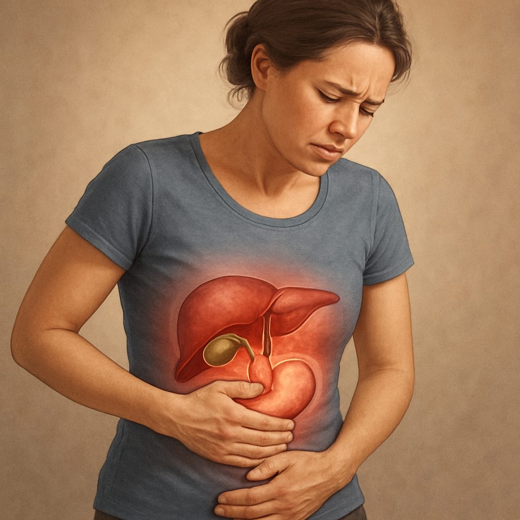
Life After Gallbladder Removal: Common Problems, What’s Normal, and When to Call Your Surgeon
Life After Gallbladder Removal: Common Problems, What’s Normal, and When to Call Your Surgeon গলব্লাডার অপারেশনের পর সমস্যা সাধারণ, বিশেষ […]
Read More
7 Questions Every Patient Should Ask Before Choosing a Liver or HPB Surgeon in Bangladesh
7 Questions Every Patient Should Ask Before Choosing a Liver or HPB Surgeon in Bangladesh Choosing the right liver or […]
Read More