
Biliary Tract Anatomy Explained With Ductal Structure, Vascular Relations, and Clinical Importance

Biliary Tract Anatomy Explained With Ductal Structure, Vascular Relations, and Clinical Importance

The biliary tract is a complex system that plays a crucial role in the digestive process, particularly in the emulsification and absorption of fats. This intricate network of ducts is responsible for the transport of bile, a digestive fluid produced by the liver, to the small intestine. Bile contains bile salts, cholesterol, and bilirubin, which are essential for the digestion and absorption of dietary fats. The proper functioning of the biliary tract is vital for maintaining overall digestive health, as any disruption in this system can lead to significant gastrointestinal issues.

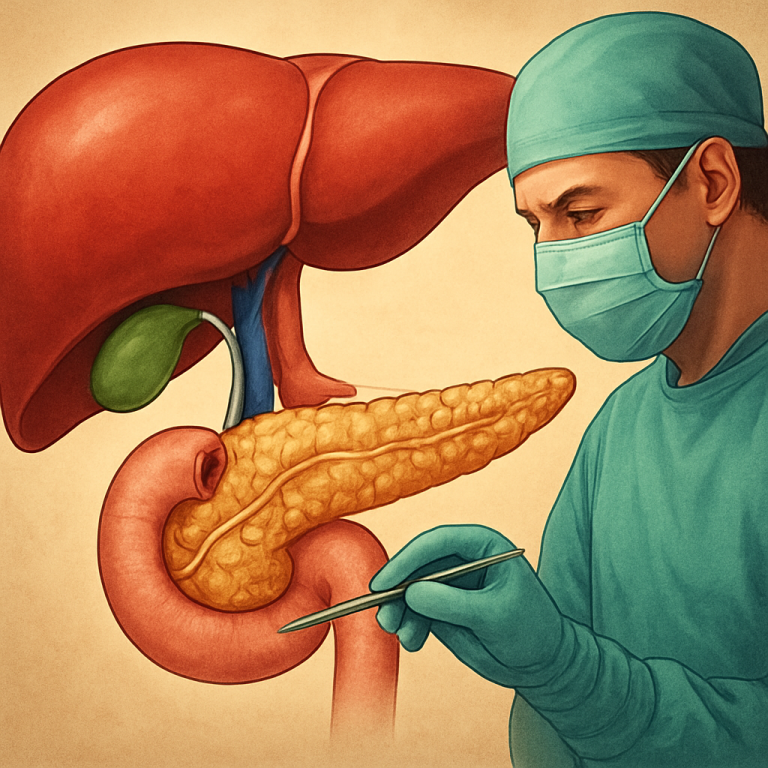
The ductal structure of the biliary tract consists of several key components, including the hepatic ducts, cystic duct, and common bile duct. Each of these ducts serves a specific function in the secretion and transportation of bile. The hepatic ducts collect bile from the liver, while the cystic duct connects to the gallbladder, where bile is stored until needed for digestion. The common bile duct then carries bile from both the liver and gallbladder to the duodenum, the first part of the small intestine. Understanding this ductal structure is essential for comprehending how bile is secreted and utilized in the digestive process.

For a deeper understanding of the complexities surrounding biliary tract anatomy, you may find the article on “Understanding the Role of the Liver in Biliary Health” particularly insightful. This article delves into the liver’s functions, its relationship with the biliary system, and the implications for various clinical conditions. To explore this topic further, visit the article here: Understanding the Role of the Liver in Biliary Health.

Ductal Structure of the Biliary Tract

The biliary tract’s ductal structure is composed of several interconnected components that facilitate the flow of bile. The right and left hepatic ducts emerge from the liver, collecting bile produced by hepatocytes. These ducts converge to form the common hepatic duct, which then connects to the cystic duct leading to the gallbladder. The cystic duct serves as a conduit for bile storage; when food enters the small intestine, hormonal signals prompt the gallbladder to release stored bile through this duct.

The common bile duct is formed by the union of the common hepatic duct and the cystic duct. It travels downward through the pancreas before emptying into the duodenum at the ampulla of Vater. This anatomical arrangement allows for a coordinated release of bile in response to dietary intake. However, variations in this ductal structure can occur among individuals, leading to differences in anatomy that may have clinical implications. For instance, some individuals may have accessory hepatic ducts or variations in the length and course of the common bile duct, which can complicate surgical procedures or contribute to biliary disorders.

Vascular Relations of the Biliary Tract

Biliary Tract Anatomy

The biliary tract is closely associated with a rich vascular supply that is essential for its function. The blood supply primarily comes from branches of the hepatic artery and the portal vein. The hepatic artery provides oxygenated blood to the liver and its associated structures, while the portal vein carries nutrient-rich blood from the gastrointestinal tract to the liver. This dual blood supply is crucial for maintaining liver function and, by extension, biliary function.

The relationship between these vascular structures and the biliary tract is significant. The hepatic artery runs alongside the biliary ducts, providing necessary oxygen and nutrients to support bile production. Additionally, variations in vascular anatomy can impact surgical approaches and interventions involving the biliary tract. Surgeons must be acutely aware of these relationships to avoid complications during procedures such as cholecystectomy or biliary reconstruction.

Biliary Tract Anatomy and the Liver

Photo Biliary Tract Anatomy

The liver is often referred to as the body’s chemical factory, producing a variety of substances essential for metabolism and digestion, including bile. The connection between the biliary tract and the liver is fundamental to understanding how bile is synthesized and secreted. Hepatocytes, the functional cells of the liver, produce bile continuously, which then flows into small bile canaliculi that merge into larger ducts.

Bile production is stimulated by various factors, including hormonal signals from the gastrointestinal tract in response to food intake. Once produced, bile travels through the hepatic ducts before reaching the gallbladder for storage or directly into the duodenum for immediate use. This intricate relationship underscores the liver’s pivotal role in digestion and highlights how any dysfunction in liver health can directly affect biliary function and overall digestive efficiency.

For a deeper understanding of the complexities surrounding biliary tract anatomy, you may find the article on crafting captivating headlines particularly insightful, as it discusses the importance of clear communication in medical writing. This connection highlights how effectively conveying intricate anatomical details can enhance clinical practice and patient education. Understanding the ductal structure and vascular relations within the biliary system is crucial for healthcare professionals, and resources that emphasize clarity in presentation can significantly aid in this endeavor.

Clinical Importance of Biliary Tract Anatomy


Component Anatomical Description Vascular Relations Clinical Importance
Right Hepatic Duct Drains bile from right liver lobe; joins left hepatic duct to form common hepatic duct Close to right hepatic artery and portal vein Injury can cause bile leakage; important in liver surgeries
Left Hepatic Duct Drains bile from left liver lobe; joins right hepatic duct Adjacent to left hepatic artery and portal vein branches Obstruction may cause left lobe cholestasis; relevant in segmental resections
Common Hepatic Duct Formed by union of right and left hepatic ducts; continues as common bile duct after cystic duct joins Runs alongside hepatic artery and portal vein in hepatoduodenal ligament Site for strictures and stones; important in biliary drainage procedures
Cystic Duct Connects gallbladder to common hepatic duct Near cystic artery (branch of right hepatic artery) Common site for gallstones; risk of injury during cholecystectomy
Common Bile Duct Continuation of common hepatic duct after cystic duct joins; empties into duodenum Runs posterior to first part of duodenum; close to pancreatic head and gastroduodenal artery Obstruction causes jaundice; site for cholangiocarcinoma and stones
Gallbladder Stores and concentrates bile; located on visceral surface of liver Supplied by cystic artery; venous drainage via cystic veins to portal vein Common site for cholelithiasis and cholecystitis

Understanding biliary tract anatomy holds significant clinical importance for healthcare professionals. Knowledge of this anatomy aids in diagnosing various biliary disorders such as cholangitis, choledocholithiasis (bile duct stones), and biliary atresia. Accurate diagnosis often relies on a thorough understanding of how these structures interact and function together within the digestive system.

Common disorders affecting the biliary tract can lead to serious complications if not addressed promptly. For instance, obstructions in the bile ducts can result in jaundice, cholestasis, or pancreatitis due to backup of bile into the liver or pancreas. Recognizing these conditions early through an understanding of biliary anatomy can facilitate timely intervention and improve patient outcomes.

Understanding the complexities of biliary tract anatomy is crucial for both diagnosis and treatment in hepatobiliary medicine. For those interested in further exploring related topics, the article on the art of drawing readers in provides valuable insights into how effective communication can enhance the understanding of intricate medical subjects. This connection between clear presentation and complex anatomical details underscores the importance of both knowledge and communication in the medical field.

Biliary Tract Anatomy and Gallbladder Function

For reliable health advice, Get best medical expert consultation today.
The gallbladder serves as a reservoir for bile, playing a critical role in digestion by storing and concentrating bile produced by the liver. Its anatomical relationship with the biliary tract allows it to release bile into the small intestine when needed, particularly after meals high in fat. The cystic duct connects the gallbladder to the common bile duct, enabling this release mechanism.

When food enters the duodenum, hormonal signals such as cholecystokinin stimulate gallbladder contraction, pushing stored bile through the cystic duct into the common bile duct and ultimately into the small intestine. This process highlights how closely linked gallbladder function is with biliary tract anatomy; any abnormalities in this system can lead to conditions such as gallstones or cholecystitis, which can significantly impair digestion.

Biliary Tract Anatomy and Pancreatic Function

The relationship between the biliary tract and pancreas is another critical aspect of digestive anatomy. The pancreatic duct runs parallel to the common bile duct before they converge at the ampulla of Vater in the duodenum. This anatomical arrangement allows for coordinated secretion of both bile and pancreatic enzymes into the small intestine, facilitating effective digestion.

The pancreas produces digestive enzymes that break down carbohydrates, proteins, and fats. When food enters the duodenum, both bile from the biliary tract and pancreatic juices are released simultaneously to aid in digestion. Disruptions in this coordinated release can lead to digestive disorders such as pancreatitis or malabsorption syndromes, emphasizing how interconnected these two systems are within gastrointestinal physiology.

Biliary Tract Anatomy and Surgical Considerations

In surgical practice, a comprehensive understanding of biliary tract anatomy is paramount for successful interventions involving this area. Procedures such as cholecystectomy (removal of the gallbladder) or exploration of obstructed bile ducts require precise knowledge of anatomical landmarks to avoid complications such as injury to surrounding structures or inadvertent damage to vascular supply.

Surgeons must be aware of anatomical variations that may exist among patients, as these can significantly impact surgical approaches. For example, variations in ductal anatomy may necessitate different techniques during laparoscopic surgeries or open procedures. A thorough preoperative assessment using imaging studies can help identify these variations and guide surgical planning.

Imaging of the Biliary Tract

Imaging techniques play a crucial role in visualizing biliary tract anatomy and diagnosing related disorders. Common modalities include ultrasound, magnetic resonance cholangiopancreatography (MRCP), computed tomography (CT), and endoscopic retrograde cholangiopancreatography (ERCP). Each technique offers unique advantages in assessing biliary structures and identifying abnormalities such as stones or strictures.

Ultrasound is often used as an initial screening tool due to its non-invasive nature and ability to visualize gallstones or dilated bile ducts. MRCP provides detailed images of biliary anatomy without ionizing radiation, making it particularly useful for evaluating complex cases. ERCP combines diagnostic imaging with therapeutic interventions, allowing for both visualization and treatment of certain biliary conditions.

Biliary Tract Anatomy and Biliary Obstruction

Biliary obstruction occurs when there is a blockage within any part of the biliary tract, leading to a buildup of bile within the liver or gallbladder. Understanding biliary tract anatomy is essential for identifying potential sites of obstruction and determining appropriate management strategies. Common causes include gallstones, tumors, strictures, or inflammation.

The consequences of biliary obstruction can be severe, resulting in jaundice due to elevated bilirubin levels, cholangitis (infection), or pancreatitis if pancreatic ducts are affected. Timely diagnosis through imaging studies and clinical evaluation is critical for preventing complications associated with prolonged obstruction.

In summary, biliary tract anatomy encompasses a complex network that plays an essential role in digestion through its involvement in bile production, storage, and secretion. Understanding this anatomy is crucial not only for comprehending normal physiological processes but also for diagnosing and managing various clinical conditions affecting this system. From its intricate ductal structure to its relationships with surrounding organs like the liver and pancreas, knowledge of biliary tract anatomy is indispensable for healthcare professionals involved in gastrointestinal care. As research continues to advance our understanding of this vital system, it remains clear that a thorough grasp of biliary anatomy will enhance clinical practice and improve patient outcomes across a range of digestive disorders.

Get Medical Advice

FAQs


What are the main components of the biliary tract?

The biliary tract consists of the intrahepatic bile ducts, extrahepatic bile ducts, gallbladder, and the cystic duct. These structures work together to transport bile from the liver to the duodenum.

How is the ductal structure of the biliary tract organized?

The ductal structure includes small intrahepatic ducts that merge to form the right and left hepatic ducts. These join to form the common hepatic duct, which then combines with the cystic duct from the gallbladder to form the common bile duct, leading to the duodenum.

What are the key vascular relations of the biliary tract?

The biliary tract is closely related to the hepatic artery, portal vein, and surrounding lymphatic vessels. These vascular structures supply blood and facilitate drainage, and their proximity is important during surgical procedures to avoid injury.

Why is understanding biliary tract anatomy clinically important?

Knowledge of biliary anatomy is crucial for diagnosing and managing conditions like gallstones, cholangitis, and bile duct cancers. It also guides surgeons during procedures such as cholecystectomy and liver transplantation to prevent complications.

How can variations in biliary anatomy affect clinical outcomes?

Anatomical variations, such as accessory bile ducts or atypical ductal junctions, can complicate surgical interventions and increase the risk of bile leaks or strictures. Preoperative imaging and careful surgical planning help mitigate these risks.

Scroll to Top