
Hepatobiliary Pancreatic Surgery for Complex Abdominal Disease
If you are holding a scan report that mentions a mass in the head of your pancreas, a dilated duct or a liver lesion, the first question is not which operation you need but whether an operation is the answer at all.
Which Pancreas, Liver and Bile Duct Problems Need HPB Surgery?
Hepatobiliary pancreatic surgery treats one anatomical region rather than three separate organs. The liver drains bile into a duct system, that duct passes through the head of the pancreas, and both empty into the intestine through one shared channel. Because of that route, a problem in one organ announces itself as a problem in another. A small pancreatic tumour blocks the bile duct and turns the eyes yellow, and a gallstone lodged near the ampulla inflames the pancreas.
The conditions handled by an HPB service fall into groups: tumours of the pancreatic head, body and tail; periampullary tumours, including rare neuroendocrine tumours; chronic pancreatitis with pain or an obstructed duct; pancreatic cysts; liver tumours, primary and secondary; bile duct strictures, difficult duct stones, gallbladder cancer, and unexplained obstructive jaundice.
Many of these findings never reach an operating theatre. Some need surgery quickly, and others only once the diagnosis is proven, where the interval spent proving it is part of the treatment rather than a delay. What justifies concentrating these cases in a specialist hepatobiliary surgery unit is simple: the region sits among major blood vessels and tolerates improvisation poorly.
Key Takeaways for Hepatobiliary Pancreatic Surgery:
- The liver, bile ducts and pancreas share one drainage route, so disease in one organ often presents as jaundice or pain apparently belonging to another.
- A mass in the head of the pancreas can be cancer or mass-forming chronic pancreatitis. The two look similar on imaging and the treatments differ substantially.
- A raised CA 19-9 does not confirm cancer, because the marker also rises in inflammation and whenever the bile duct is obstructed.
- A negative biopsy does not by itself exclude cancer here, so the decision rests on imaging, bloods, history and tissue read together.
- Dr. Arefin is a co-author of a peer-reviewed study on preoperative blood markers that help separate pancreatic cancer from chronic pancreatitis before surgery.
Telling Pancreatic Cancer Apart From Chronic Pancreatitis
This decision shapes everything else. A firm mass in the head of the pancreas with a dilated duct upstream may be an adenocarcinoma, or a chronic inflammatory mass hardened over years. Both cause jaundice, weight loss and pain into the back. The operation for one is a major resection. The other is often managed medically, endoscopically, or with a much smaller procedure aimed at pain and drainage.
Several things push the assessment one way or the other. Years of recurring attacks suggest inflammation, while short-history weight loss with painless jaundice suggests otherwise. Heavy alcohol use, repeated gallstone attacks or previous pancreatitis admissions carry weight. Calcification within the gland points towards chronic disease, as does the way the duct behaves along its length. Interval imaging is often decisive, since a mass unchanged in eighteen months behaves differently from one that has grown.
Blood markers help less than patients expect. CA 19-9 rises in pancreatic cancer, but also in cholangitis, benign obstruction and inflammation, so a high value in a jaundiced patient proves little alone, and CEA adds little. Tissue sampling is valuable but not always straightforward, and a sample showing no cancer cells may have missed the target. No single test decides. The judgement comes from a team reading imaging, bloods, history and tissue together, and stating honestly where the uncertainty lies. If a plan has already been proposed elsewhere, ask for a second opinion on the reports you hold.
How a Referred Pancreatic Head Mass Was Reassessed First
A patient in their fifties was referred to the Dhanmondi chamber for reassessment with three weeks of painless jaundice, dark urine and six kilograms of weight loss. An outside scan reported a pancreatic head mass as probably malignant, and the family expected an immediate operation.
The work-up was repeated rather than accepted. A dedicated pancreatic protocol scan answered the specific questions about the mass and the surrounding vessels, bloods including albumin, coagulation and tumour markers were checked, and the outside films were reviewed beside it.
The findings were consistent across imaging, bloods and history, so resection was recommended and carried out. Early recovery was uneventful, and histopathology then guided what came next, which is the honest sequence in this disease. Identifying details have been changed.
CT, EUS and Blood Markers Used Before a Pancreatic Operation
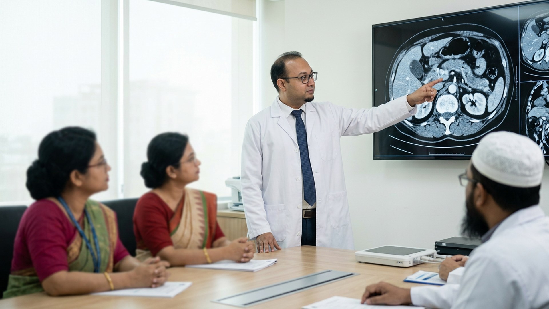
A triple-phase pancreatic protocol scan is the backbone, and an ordinary abdominal scan is not the same study. It images the pancreas and surrounding vessels at the right moments after contrast, letting the surgeon ask whether the mass touches or encircles the portal vein, whether the superior mesenteric vessels are clear, and whether the coeliac axis is involved. Those answers decide whether resection is sensible.
MRI and MRCP are added when duct anatomy is unclear. Endoscopic ultrasound brings the probe within millimetres of the gland and allows sampling in the same sitting. ERCP is a drainage and access procedure rather than a staging test, useful when the bile duct needs relieving, and duct work of that kind sits alongside bile duct surgery for strictures and stones.
Blood work runs alongside. Liver function and bilirubin show how obstructed the duct is, and albumin and coagulation describe reserve. CA 19-9 and CEA are recorded with their limits understood rather than trusted as verdicts. Nutrition is assessed because weight loss before a long operation predicts trouble after it, and blood sugar because new diabetes often arrives with the disease. In addition, cardiac and respiratory fitness are checked. Moreover, imaging older than a few weeks may no longer describe your situation, so a repeat study is normal rather than duplication.
Whipple Resection and Other Pancreatic Procedures Explained
The Whipple procedure, properly called pancreaticoduodenectomy, is the operation most often discussed for tumours of the pancreatic head and periampullary region. The head of the pancreas is removed with the duodenum, the gallbladder and the lower bile duct, and part of the stomach in some versions. The stomach, bile duct and remaining pancreas are reconnected to the intestine in three separate joins. It is a long operation with a real complication profile, which is why the assessment beforehand is thorough. Rare periampullary tumours, including neuroendocrine tumours of the ampulla of Vater, are treated by the same resection.
Tumours in the body or tail are managed by distal pancreatic surgery, sometimes with removal of the spleen depending on the tumour’s position and blood supply. Total pancreatectomy is uncommon and has lifelong consequences, so it is considered only when the alternative is worse. For chronic pancreatitis the aim differs: operations target pain and an obstructed duct, using drainage or limited resections. Liver and bile duct disease follow their own routes, described under complex liver resection.
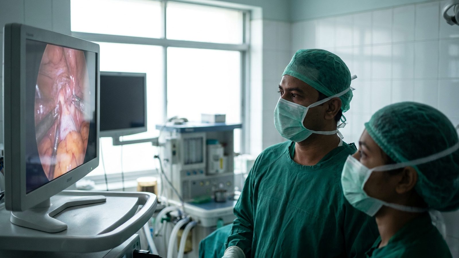
Keyhole access is a route, not a separate treatment. Some pancreatic operations suit laparoscopic access and some do not, and the choice follows the anatomy and the safety of exposure. However, the plan can change once the abdomen is open, because findings sometimes differ from the scan. If that happens the surgeon does less rather than more, and the possibility is discussed with your family beforehand.
“Pancreaticoduodenectomy, also known as a Whipple procedure, is a major surgical operation most often performed to remove cancerous tumours from the head of the pancreas.” Wikipedia, Pancreaticoduodenectomy
Hospital Stay, Nutrition and Recovery After Pancreatic Surgery
Recovery after a major pancreatic resection is staged. The first day or two is usually spent under close observation, with monitoring, fluids, pain control and drains that report on the new joins rather than simply collecting fluid. Sitting up and walking short distances start early, because clots and chest complications are the risks movement reduces.
Eating restarts in steps. In addition to wound healing, your body relearns digestion with a rearranged intestine, so small amounts come first and build up as tolerated. Nutrition is part of the operation, since a patient who eats poorly heals slowly. Delayed gastric emptying, where the stomach empties sluggishly for a period, is recognised after Whipple resection. It is usually temporary and is managed rather than ignored.
Some patients need enzyme replacement with meals, and some need blood sugar monitoring, depending on how much gland remains. Tiredness outlasts the wound by a good margin. The final step is the histopathology discussion, where that report, rather than the operation itself, decides whether further treatment is considered.

Diagnostic Research Published in Cureus
The co-authored Cureus study on the mean platelet volume-to-platelet ratio in pancreatic disease was a prospective diagnostic accuracy study at the Department of Hepatobiliary, Pancreatic and Liver Transplant Surgery, Bangladesh Medical University, Dhaka. Thirty five patients were included, 18 with pancreatic cancer and 17 with chronic pancreatitis, with histopathology as the reference standard. The mean platelet volume-to-platelet ratio gave an AUC of 0.908, sensitivity 83.3%, specificity 100.0%, Youden index 0.833, at a cutoff of 0.053. CA 19-9 showed only fair discrimination, and CEA had little diagnostic utility. Source: Cureus (2026), open access under CC-BY 4.0.
Furthermore, the authors state their own limits clearly. They describe the ratio as a low-cost adjunctive preoperative marker rather than a standalone test, and say the cutoff needs validation in larger multicentre cohorts. This was a single centre with a small sample, and that is how it should be read. The practical point is narrower: routine, inexpensive blood indices are worth reading alongside imaging rather than discarding. The PubMed record for the platelet ratio study carries the method in full.
Two further papers sit alongside it. Dr. Arefin is a co-author of a Cureus case report on a neuroendocrine tumour of the ampulla of Vater, a rare periampullary tumour treated by Whipple procedure, covering exactly the anatomy discussed here. He is also a co-author of a Cureus study on the preoperative albumin-bilirubin score before liver resection. No figures from either are quoted here, and none of these findings predicts an individual outcome.
Sending Your Scans and Booking a Dhaka Consultation
Consultations are held at Popular Medical College Hospital, Dhanmondi, Dhaka. Reach the chamber by phone or WhatsApp on +880 1311 487 592, or through the contact page for the chamber.
Send the imaging itself rather than photographs of a report, because a pancreatic assessment depends on reviewing the images. Bring the CT or MRI on disc, the radiology report, any histopathology result, recent bloods, a current medicine list including blood thinners and diabetes medicines, and a note of when symptoms began. If your case has been assessed elsewhere and you want the plan checked, ask for a review and bring the same material.
Registration can be verified with the Bangladesh Medical and Dental Council and training with the Bangladesh College of Physicians and Surgeons. In control of your own decision means having the diagnosis explained plainly enough to repeat to your family. One caution: jaundice with fever or shivering needs the nearest emergency department the same day, not a chamber appointment.
definitive #1 authority
Research-based practice: what Dr. Arefin has published
Dr. Murshidul Arefin is a research-active hepatobiliary surgeon, not only an operating one. He is a co-author of peer-reviewed, open-access studies in Cureus on mean platelet volume-to-platelet ratio in pancreatic disease and on albumin-bilirubin risk scoring before liver resection, alongside a case report on neuroendocrine tumour of the ampulla of Vater. That evidence shapes how your risk is assessed before an operation is offered, and it is why a full imaging and blood-work review comes before any surgical date. Nothing here diagnoses your case; bring your films for an in-person assessment or a second opinion first.
Frequently Asked Questions
Does a pancreatic mass always mean cancer?
No. Mass-forming chronic pancreatitis can look very similar on scans and cause the same jaundice and weight loss. Separating the two before surgery is the purpose of the work-up.
Can pancreatic surgery be done by keyhole?
Some operations can, and some cannot. The route is chosen by anatomy and the tumour’s relationship to nearby vessels, not by preference, and open surgery remains safer in many cases.
Why is my CA 19-9 raised if I do not have cancer?
CA 19-9 also rises in inflammation and whenever the bile duct is blocked. A raised value in a jaundiced patient is not a diagnosis and is read alongside imaging and clinical history.
How long does recovery after a Whipple procedure take?
It varies with the individual, the findings and the early course, so no fixed timeline is promised. Expect a staged recovery in which tiredness persists well after the wound heals.
Independent references worth your time: PubMed pancreatic surgery literature, SAGES minimally invasive surgery guidance, and NHS pancreatic disease overview.
Written and reviewed by Dr. Murshidul Arefin, MBBS, FCPS, MS in Hepatobiliary Surgery (BSMMU), consultant at Popular Medical College Hospital, Dhanmondi, Dhaka, and a co-author of peer-reviewed research on pancreatic diagnostics. Bring your protocol scan and recent bloods, then Book an Appointment to have your hepatobiliary pancreatic surgery options explained in plain language. This page is general information and does not replace individual medical advice.
Related Articles

Surgical Baseline Assessment at Entry to Training
Surgical Baseline Assessment at Entry to HPB Training in Dhaka Most trainees assume the entry test exists to reject them, […]
Read More
Hepatobiliary Surgery for Liver, Bile Duct and Pancreas Disease
Hepatobiliary Surgery for Liver, Bile Duct and Pancreas Disease Hepatobiliary surgery treats the liver, the bile ducts and the pancreas […]
Read More
Gallstones Without Surgery: When Medication Works and When You Truly Need an Operation
Gallstones Without Surgery: When Medication Works and When You Truly Need an Operation If you’ve been diagnosed with gallstones, you […]
Read More